
Page 119 - Read Online
P. 119
distal root of the third molar and retromolar area, and muscles. The pain impulses may be generated upon
this distribution showed that the contents of this canal compression or stimulation of the plexus and be carried
innervate and supplied the third molar and mucosa of the away causing pain escape in the presence of a complete
retromolar area. nerve block [Figure 1].
[14]
Coleman and Smith speculated that aberrant nerve
branches to the mandibular teeth and periodontium DISCUSSION
arising from major branches of the mandibular trunk high
within the pterygomandibular space could also be bathed Since the early 1970s, dentistry has experienced a
by anesthetic deposited at the mandibular neck. These resurgence of interest in the neuro-anatomical basis of
branches would probably escape the drug when it is local anesthesia, resulting in many scientific reports on
[6,17]
[14]
deposited at the mandibular foramen. The authors also the subject. Numerous studies have provided a detailed
[15]
[16]
cited Sutton’s and Rood’s papers which suggested knowledge of the anatomy of the trigeminal nerve, which
[18-21]
that there may be accessory innervation of the mandibular is important in obtaining profound local anesthesia.
teeth from branches of the lingual, buccal, facial, and To explain the incidence of inadequate anesthesia in the
mandibular region despite an efficient inferior alveolar
upper cervical nerves from their clinical experience. With
the exception of the buccal nerve, there is little anatomic nerve block, an EPP was first described by Carter and
[1]
evidence to support these opinions. Keen. It was suggested to deposit local anesthetic
solution in the vicinity of the retromandibular foramen
Neurovascular plexus theory to prevent the pain escape. However, the persistence of
[6]
The above authors have demonstrated “accessory” nerves pain escape noted even after infiltrating the retromolar
from the lateral pterygoid muscle, the temporal muscle, area with lidocaine solution in 5 of our cases. Recently,
the auriculotemporal nerve, and the mylohyoid nerve. Ngeow noted the incidence of EPP while the elevation
[2]
In most instances, these accessory nerves pass through of an impacted tooth and assumed it as a result of
foramina of the condylar neck, retromolar fossa, or within compression of inferior alveolar nerve. It was postulated
the infratemporal fossa to form a neural plexus which that the release of sodium and potassium ions from the
communicates with the inferior alveolar neurovascular compressed nerve may be responsible for propagating the
bundle. However, all of the authors note that this accessory pain impulses. This hypothesis was criticized because
[2]
nerve or the plexus innervates the third molar. Conceptually, the pressure would result in paresthesia which sustains
if this nerve plexus does, in fact, supply the third molar, long even after the procedure.
then the pain would be expected from the commencement The theory based on present literature validated the
of tooth removal procedure, and not specifically during presence of a plexus at the apical region of the tooth
elevation of the tooth or socket curettage. which may be stimulated by inadvertent tooth elevation
Based on the current literature search, the authors or postextraction curettage.
hypothesize that the EPP in lower third molar surgery The incidence of pain escape seems to occur only
can be attributed to the occasional presence of a NVP during third molar surgery because of the inclination
lying deep to the roots of the third molar which does impacted tooth, as well as the curvature of the angle of
not provide innervation. This plexus may be formed by the mandible. This brings the neurovascular bundle in
various nerves including the auriculotemporal, mylohyoid, proximity to the tooth root. It is not seen in the first
and retromolar plexus from the pterygoid and masseter and second molar regions as there is no possibility of
compression of the neurovascular bundle.
There was also a significant incidence of bleeding
following the EPP, which usually noted immediately
following the elevation of the tooth fragment. This may
be secondary to damage inferior alveolar vessels or
vessels from the NVP.
a A large-scale cadaveric study would confirm the presence
b of these independent NVP and their third molar
innervations.
d c REFERENCES
1. Carter RB, Keen EN. The intramandibular course of the inferior alveolar
nerve. J Anat 1971;108:433‑40.
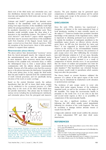
Figure 1: Diagrammatic illustration of escape pain phenomenon (EPP)
and neurovascular plexus (NVP) theory during the two section technique 2. Ngeow WC. Tooth section technique for wisdom teeth. Int J Oral Maxillofac Surg
of third molar removal (inner figure: mesioangular mandibular third 2009;38:908.
molar impaction). (a) Conduction block; (b) neurovascular/nerve plexus 3. Arakeri G, Arali V. Tooth section technique and pain upon elevation in third
entering into the bony canal; (c) compression of the neurovascular molar removal. Int J Oral Maxillofac Surg 2010;39:98‑9.
bundle; (d) escape of pain away from conduction blockade through 4. Anil A, Peker T, Turgut HB, Gülekon IN, Liman F. Variations in the anatomy
neurovascular/nerve plexus of the inferior alveolar nerve. Br J Oral Maxillofac Surg 2003;41:236‑9.
Plast Aesthet Res || Vol 2 || Issue 3 || May 15, 2015 109