
Page 116 - Read Online
P. 116
printed condyle/fossa reconstruction in adults with
ankylosis [Figure 24] with consideration for integration
[18]
in the facial rotation procedure for hemifacial microsomia
following rib graft failure.
This article represents level 5 evidence, and therefore
a c e g i simply provides an expert opinion. The variability of
pathology, lack of a gold standard, different surgical
experiences, duration of the phased treatment, desiderata,
compliance and economic situation of the patients, and
use of new technologies prohibit valid sampling and
prospective analyses. Nonetheless, it is hoped that the
b d f h j comprehensive treatment planning described in this
Figure 22: The same patient as in Figure 20. (a and b) Before joint report may be used to promote optimal patient care.
reconstruction; (c and d) after joint reconstruction; (e and f) before facial
rotation, after orthodontic preparation; (g and h) after facial rotation;
(i and j) after three-dimensional (3D) titanium print implantation of the REFERENCES
right mandible
1. Grabb WC. The first and second branchial arch syndrome. Plast Reconstr
Surg 1965;36:485‑508.
2. Vento AR, LaBrie RA, Mulliken JB. The O.M.E.N.S. classification of hemifacial
microsomia. Cleft Palate Craniofac J 1991;28:68‑76.
3. Pruzansky S. Not all dwarfed mandibles are alike. Birth Defects 1969;5:120‑9.
4. Kaban LB, Moses MH, Mulliken JB. Surgical correction of hemifacial
microsomia in the growing child. Plast Reconstr Surg 1988;82:9‑19.
5. Harvold EP, Vargervik K, Chirici G. Treatment of Hemifacial Microsomia.
New York: Alan R. Liss; 1983.
6. Kaban LB, Moses MH, Mulliken JB. Correction of hemifacial microsomia in
a b the growing child: a follow‑up study. Cleft Palate J 1986;23 Suppl 1:50‑2.
7. Mommaerts MY, Nagy K. Is early osteodistraction a solution for the
ascending ramus compartment in hemifacial microsomia? A literature study.
J Craniomaxillofac Surg 2002;30:201‑7.
8. Nagy K, Kuijpers‑Jagtman AM, Mommaerts MY. No evidence for long‑term
effectiveness of early osteodistraction in hemifacial microsomia. Plast Reconstr
Surg 2009;124:2061‑71.
9. Obwegeser HL. Correction of the skeletal anomalies of oto‑mandibular
dysostosis. J Maxillofac Surg 1974;2:73‑92.
10. Peltomäki T. Growth of the costochondral junction and its potential
c d applicability for the reconstruction of the mandibular condyle. Turku:
Turunyliopisto; 1993.
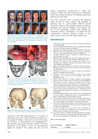
Figure 23: Pruzansky-Kaban Type III. Microvascular fibula transfer in 11. Molina F, Ortiz Monasterio F. Mandibular elongation and remodeling by
a 10-year-old patient. (a) Harvesting of the fibula; (b) microvascular distraction: a farewell to major osteotomies. Plast Reconstr Surg 1995;96:825‑40.
anastomosis; (c) three-dimensional (3D) computed tomography (CT) 12. Ortiz Monasterio F, Molina F, Andrade L, Rodriguez C, Sainz Arregui J.
reconstruction after fibula transplantation to the left mandible; (d) 3D Simultaneous mandibular and maxillary distraction in hemifacial microsomia
CT reconstruction, profile view in adults: avoiding occlusal disasters. Plast Reconstr Surg 1997;100:852‑61.
13. Kunz C, Brauchli L, Moehle T, Rahn B, Hammer B. Theoretical considerations
for the surgical correction of mandibular deformity in hemifacial microsomia
patients using multifocal distraction osteogenesis. J Oral Maxillofac Surg
2003;61:364‑8.
14. Stelnicki EJ, Boyd JB, Nott RL, Barnavon Y, Uecker C, Henson T. Early treatment
of severe mandibular hypoplasia with distraction mesenchymogenesis and
bilateral free fibula flaps. J Craniofac Surg 2001;12:337‑48.
15. Lee SJ, Lee HP, Tse KM, Cheong EC, Lim SP. Computer‑aided design and
rapid prototyping‑assisted contouring of costal cartilage graft for facial
reconstructive surgery. Craniomaxillofac Trauma Reconstr 2012;5:75‑82.
16. Tanna N, Broer PN, Roostaeian J, Bradley JP, Levine JP, Saadeh PB. Soft tissue
correction of craniofacial microsomia and progressive hemifacial atrophy.
J Craniofac Surg 2012;23:2024‑7.
a b 17. Lim AA, Fan K, Allam KA, Wan D, Tabit C, Liao E, Kawamoto HK,
Bradley JP. Autologous fat transplantation in the craniofacial patient: the
Figure 24: Bilateral three-dimensional printed temporomandibular joint (TMJ)
prostheses. (a) TMJ ankylosis present; (b) fossa/condyle prosthesis in place UCLA experience. J Craniofac Surg 2012;23:1061‑6.
18. Haq J, Patel N, Weimer K, Matthews NS. Single stage treatment of ankylosis
of the temporomandibular joint using patient‑specific total joint replacement
popularity of 3D early osteodistraction, [11,12] and then its and virtual surgical planning. Br J Oral Maxillofac Surg 2014;52:350‑5.
subsequent fall in use. Microvascular fibula transfer in a
[13]
young patient [Figure 23] has been used, but the author How to cite this article: Mommaerts MY. Hemifacial microsomia:
[14]
has returned to nonvascularized rib transplantation. management of the vertical ramus compartment. Plast Aesthet Res
[15]
2015;2:99-106.
Microvascular parascapular dermofat transfer has been
[16]
abandoned by the author in favor of nonvascularized Source of Support: Nil, Conflict of Interest: None declared.
gluteal fat transfer. The author has also used 3D Received: 01-02-2015; Accepted: 07-04-2015
[17]
106 Plast Aesthet Res || Vol 2 || Issue 3 || May 15, 2015