
Page 112 - Read Online
P. 112
a b c d e
a b c
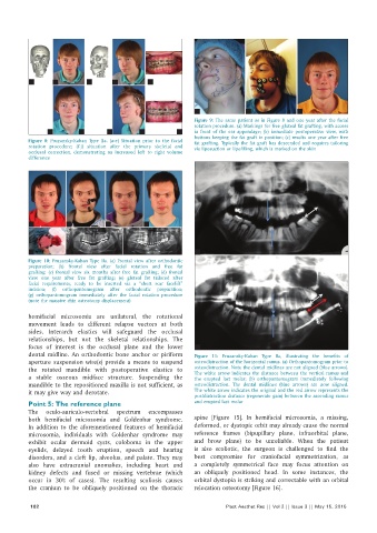
Figure 9: The same patient as in Figure 8 and one year after the facial
rotation procedure. (a) Markings for free gluteal fat grafting, with access
f g h i j in front of the ear appendage; (b) immediate postoperative view, with
buttons keeping the fat graft in position; (c) results one year after free
Figure 8: Pruzansky-Kaban Type IIa. (a-e) Situation prior to the facial fat grafting. Typically the fat graft has descended and requires tailoring
rotation procedure; (f-j) situation after the primary skeletal and via liposuction or lipofilling, which is marked on the skin
occlusal correction, demonstrating an increased left to right volume
difference
a b c d
e f g a
Figure 10: Pruzansky-Kaban Type IIa. (a) Frontal view after orthodontic
preparation; (b) frontal view after facial rotation and free fat
grafting; (c) frontal view six months after free fat grafting; (d) frontal
view one year after free fat grafting; (e) gluteal fat tailored after
facial requirements, ready to be inserted via a “short scar facelift”
incision; (f) orthopantomogram after orthodontic preparation;
(g) orthopantomogram immediately after the facial rotation procedure
(note the massive chin osteotomy displacement)
hemifacial microsomia are unilateral, the rotational
movement leads to different relapse vectors at both
sides. Interarch elastics will safeguard the occlusal
relationships, but not the skeletal relationships. The
focus of interest is the occlusal plane and the lower b
dental midline. An orthodontic bone anchor or piriform Figure 11: Pruzansky-Kaban Type IIa, illustrating the benefits of
aperture suspension wire(s) provide a means to suspend osteodistraction of the horizontal ramus. (a) Orthopantomogram prior to
the rotated mandible with postoperative elastics to osteodistraction. Note the dental midlines are not aligned (blue arrows).
The white arrow indicates the distance between the vertical ramus and
a stable osseous midface structure. Suspending the the erupted last molar; (b) orthopantomogram immediately following
mandible to the repositioned maxilla is not sufficient, as osteodistraction. The dental midlines (blue arrows) are now aligned.
it may give way and derotate. The white arrow indicates the original and the red arrow represents the
postdistraction distance (regenerate gain) between the ascending ramus
Point 5: The reference plane and erupted last molar
The oculo-auriculo-vertebral spectrum encompasses
both hemifacial microsomia and Goldenhar syndrome. spine [Figure 15]. In hemifacial microsomia, a missing,
In addition to the aforementioned features of hemifacial deformed, or dystopic orbit may already cause the normal
microsomia, individuals with Goldenhar syndrome may reference frames (bipupillary plane, infraorbital plane,
exhibit ocular dermoid cysts, coloboma in the upper and brow plane) to be unreliable. When the patient
eyelids, delayed tooth eruption, speech and hearing is also scoliotic, the surgeon is challenged to find the
disorders, and a cleft lip, alveolus, and palate. They may best compromise for craniofacial symmetrization, as
also have extracranial anomalies, including heart and a completely symmetrical face may focus attention on
kidney defects and fused or missing vertebrae (which an obliquely positioned head. In some instances, the
occur in 30% of cases). The resulting scoliosis causes orbital dystopia is striking and correctable with an orbital
the cranium to be obliquely positioned on the thoracic relocation osteotomy [Figure 16].
102 Plast Aesthet Res || Vol 2 || Issue 3 || May 15, 2015