
Page 111 - Read Online
P. 111
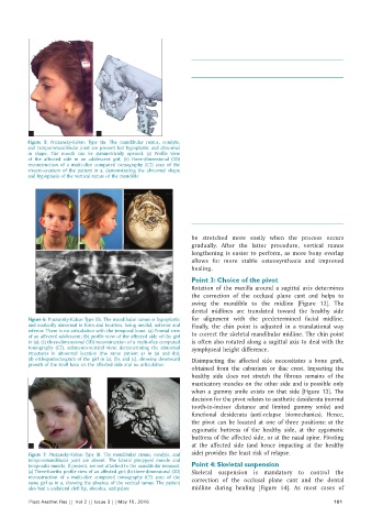
Table 1: General treatment strategies based on
                                                              the Pruzansky‑Kaban classification of mandibular
                                                              abnormalities
                                                               Pruzansky-Kaban   Treatment strategies
                                                               type
                                                               Type I           Orthognathic surgical correction “facial
                                                                                rotation”  after orthodontic alignment,
                                                                                     [9]
                                                                                coordination and decompensation. Standard
                                                                                le Fort I, bilateral sagittal split osteotomies,
                                                                                and sliding genioplasty techniques are used
                                                               Type IIa         Surgery is only performed early at the
                                                                                age of 4 and older, when there is a centric
                                                                                occlusion-centric relation shift of more
                                                                                than 5 mm. The surgery involves joint
           a                    b                                               reconstruction with costochondral grafting
          Figure 5:  Pruzansky-Kaban Type  IIa.  The  mandibular  ramus,  condyle,   Osteodistraction in the horizontal (not
          and temporomandibular joint are present but hypoplastic and abnormal   vertical) ramus is performed when there is
          in shape. The mouth can be symmetrically opened. (a) Profile view     insufficient bone stock to perform a sagittal
          of the affected side in an adolescent  girl; (b) three-dimensional (3D)   split osteotomy after puberty
          reconstruction of a  multi-slice  computed  tomography (CT) scan of the
          viscero-cranium of the patient in a, demonstrating the abnormal shape   Orthognathic surgical correction “facial
          and hypoplasia of the vertical ramus of the mandible                  rotation” after orthodontic alignment,
                                                                                coordination, and decompensation is
                                                                                performed at puberty and later
                                                               Type IIb and III  Joint and ramus reconstruction at the age of
                                                                                4 and older. Orthognathic surgical correction
                                                                                “facial rotation” after orthodontic alignment,
                                                                                coordination and decompensation, at
                                                                                puberty and later


                                                              be stretched more easily when the process occurs
             a             b             c                    gradually. After the latter procedure, vertical ramus
                                                              lengthening is easier to perform, as more bony overlap
                                                              allows for more stable osteosynthesis and improved
                                                              healing.
                                                              Point 3: Choice of the pivot
                                                              Rotation of the maxilla around a sagittal axis determines
                                                              the correction of the occlusal  plane cant and helps to
                                                              swing  the mandible to the  midline  [Figure  12]. The
             d                                                dental midlines  are  translated toward the  healthy  side
          Figure 6: Pruzansky-Kaban Type IIb. The mandibular ramus is hypoplastic   for alignment with the predetermined facial midline.
          and markedly abnormal in form and location, being medial, anterior and   Finally,  the  chin  point is  adjusted in  a  translational way
          inferior. There is no articulation with the temporal bone. (a) Frontal view
          of an affected adolescent; (b) profile view of the affected side of the girl   to correct the skeletal mandibular midline. The chin point
          in (a); (c) three-dimensional (3D) reconstruction of a multi-slice computed   is often also rotated along a sagittal axis to deal with the
          tomography (CT), submento-vertical view,  demonstrating  the  abnormal   symphyseal height difference.
          structures in abnormal location (the same patient as in (a)  and (b));
          (d) orthopantomogram of the girl in (a), (b), and (c), showing downward   Disimpacting the affected side necessitates a bone graft,
          growth of the skull base on the affected side and no articulation
                                                              obtained from the calvarium or iliac crest. Impacting the
                                                              healthy side does not stretch the fibrous remains of the
                                                              masticatory muscles on the other side and is possible only
                                                              when a gummy smile exists on that side  [Figure  13]. The
                                                              decision for the pivot relates to aesthetic desiderata (normal
                                                              tooth-to-incisor distance and limited gummy smile) and
                                                              functional  desiderata  (anti-relapse  biomechanics).  Hence,
                                                              the pivot can be located at one of three positions: at the
                                                              zygomatic buttress of the healthy side, at the zygomatic
                                                              buttress of the affected side, or at the nasal spine. Pivoting
           a                        b                         at the affected side  (and hence impacting at the healthy
          Figure 7:  Pruzansky-Kaban Type III. The mandibular ramus, condyle, and   side) provides the least risk of relapse.
          temporomandibular  joint are absent. The lateral pterygoid  muscle  and
          temporalis muscle, if present, are not attached to the mandibular remnant.   Point 4: Skeletal suspension
          (a) Three-fourths profile view of an affected girl; (b) three-dimensional (3D)   Skeletal suspension is mandatory to control the
          reconstruction of a multi-slice computed tomography (CT) scan of the   correction of the occlusal plane cant and the dental
          same girl as in a, showing the absence of the vertical ramus. The patient
          also had a unilateral cleft lip, alveolus, and palate  midline  during  healing  [Figure  14].  As  most  cases  of
          Plast Aesthet Res || Vol 2 || Issue 3 || May 15, 2015                                             101