
Page 114 - Read Online
P. 114
a b
a
c d
b
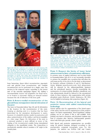
Figure 17: Pruzansky-Kaban Type IIb. Reconstruction of the
temporomandibular joint. (a) Two harvested rib segments, one
containing bony and cartilaginous components and the other containing
only cartilage; (b) the zygomatic arch has been reconstructed using
calvarial bone (two small arrows pointing at the micro osteosynthesis
screws) and the fossa has been reconstructed using the cartilaginous
segment (not visible). The condyle has been reconstructed using the
e costochondral segment, osteosynthetized via the temporal approach (big
Figure 16: Goldenhar syndrome with scoliosis and orbital dystopia. The arrow pointing at oblique osteosynthesis screws)
left orbit has been repositioned 1 cm higher. Free gluteal fat grafting,
micro lipofilling, and a face-lift on the affected side were also performed Point 9: Respect the limits of lower facial
after joint reconstruction and facial rotation. (a) Frontal view before the
aforementioned procedures; (b) frontal view after the aforementioned advancement in favor of masticatory efficiency
procedures; (c) profile view before the aforementioned procedures; In extreme cases of sagittal deficiency such as that found
(d) profile view after the aforementioned procedures; (e) intraoperative in Pruzansky-Kaban Type III it is not necessarily desirable
view of the transcranial orbital repositioning (with the assistance of
P. Staels, neurosurgeon) to advance the mandible into a position that will allow the
soft tissue profile of the chin to be the ideal, as determined
bone harvesting, donor defect reconstruction, zygomatic by Facewizz software (Orthoface R and D, Sint-Martens-
arch and glenoid fossa reconstruction, and condylar Latem, Belgium) (www.facewizz.com). Such advancement
reconstruction) can be performed via a single, wave line will be opposed by the sphenomandibular ligament
incision in the temporal region, extending to the lowest and the geniohyoid muscles, thereby jeopardizing the
part of the auricular appendage [Figure 18]. Adding a maintenance of occlusal stability. Maxillary, mandibular, and
retromandibular incision will jeopardize the facial nerve, chin advancements should be tailored to the encountered
as its location is abnormal. Adding an intraoral incision strain. In these instances, chin augmentation with calcium
increases the risk of infection of the bone graft. phosphate paste (Hydroset, Stryker, Kalamazoo, MI, USA)
can be helpful in increasing chin projection [Figure 20].
Point 8: Antero‑medial reconstruction of the
glenoid fossa versuspostero‑lateral relocation of Point 10: Reconstruction of the lateral and
the joint posterior ramus with added manufacturing
The issue in Pruzansky-Kaban Type IIb and III deformities technology
is the location for the reconstruction of the joint. Several options exist for augmentation of the lateral aspect
Creating an abutting joint in a location that has been of the ramus, but few exist for augmentation of the posterior
determined by the anomalous development is easier; aspect. For lateral augmentation, sliced lyophilized cartilage
however, it is doubtful whether medial reconstruction will grafting can be an option if this is still available. Bone
allow symmetrization of the midface and lateral mandible grafting may lead to resorption, and alloplastic implants may
at a later stage [Figure 19]. Relocation of the joint to lead to extrusion after infection, hydroxyapatite granules
a mirrored position is more difficult in terms of the mixed with fibrin glue provide a better option.
healing of the reconstructed condyle being transplanted Both lateral and posterior augmentation are possible
obliquely to the mandibular stump. The composition is using 3D printed titanium, designed according to the
mechanically unstable when it assumes a 30° angle in the postoperative computed tomography scans. For this
frontal plane. purpose, the authors use ProPlan CMF and 3-matic
104 Plast Aesthet Res || Vol 2 || Issue 3 || May 15, 2015