
Page 137 - Read Online
P. 137
Chen et al. Characteristics of viral encephalitis with epilepsy
examination is divided into five grading standards, macrophages, enhance B cell and T cell activity,
that is, normality, marginal state, mild abnormality, and slow viral replication. However, patients in my
moderate abnormality, and severe abnormality. research have not used interferon due to its potential
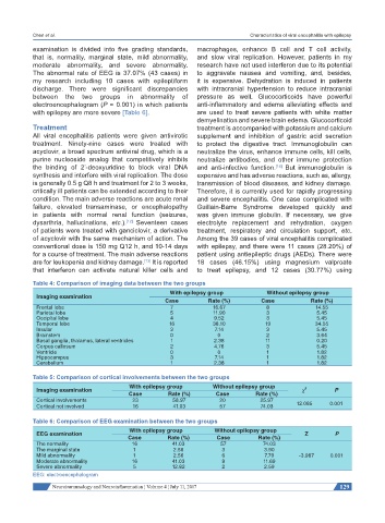
The abnormal rate of EEG is 37.07% (43 cases) in to aggravate nausea and vomiting, and, besides,
my research including 10 cases with epileptiform it is expensive. Dehydration is induced in patients
discharge. There were significant discrepancies with intracranial hypertension to reduce intracranial
between the two groups in abnormality of pressure as well. Glucocorticoids have powerful
electroencephalogram (P = 0.001) in which patients anti-inflammatory and edema alleviating effects and
with epilepsy are more severe [Table 6]. are used to treat severe patients with white matter
demyelination and severe brain edema. Glucocorticoid
Treatment treatment is accompanied with potassium and calcium
All viral encephalitis patients were given antivirotic supplement and inhibition of gastric acid secretion
treatment. Ninety-nine cases were treated with to protect the digestive tract. Immunoglobulin can
acyclovir, a broad spectrum antiviral drug, which is a neutralize the virus, enhance immune cells, kill cells,
purine nucleoside analog that competitively inhibits neutralize antibodies, and other immune protection
the binding of 2’-deoxyuridine to block viral DNA and anti-infective function. But immunoglobulin is
[14]
synthesis and interfere with viral replication. The dose expensive and has adverse reactions, such as, allergy,
is generally 0.5 g Q8 h and treatment for 2 to 3 weeks, transmission of blood diseases, and kidney damage.
critically ill patients can be extended according to their Therefore, it is currently used for rapidly progressing
condition. The main adverse reactions are acute renal and severe encephalitis. One case complicated with
failure, elevated transaminase, or encephalopathy Guillain-Barre Syndrome developed quickly and
in patients with normal renal function (seizures, was given immune globulin. If necessary, we give
dysarthria, hallucinations, etc.). Seventeen cases electrolyte replacement and rehydration, oxygen
[12]
of patients were treated with ganciclovir, a derivative treatment, respiratory and circulation support, etc.
of acyclovir with the same mechanism of action. The Among the 39 cases of viral encephalitis complicated
conventional dose is 150 mg Q12 h, and 10-14 days with epilepsy, and there were 11 cases (28.20%) of
for a course of treatment. The main adverse reactions patient using antiepileptic drugs (AEDs). There were
are for leukopenia and kidney damage. It is reported 18 cases (46.15%) using magnesium valproate
[13]
that interferon can activate natural killer cells and to treat epilepsy, and 12 cases (30.77%) using
Table 4: Comparison of imaging data between the two groups
With epilepsy group Without epilepsy group
Imaging examination
Case Rate (%) Case Rate (%)
Frontal lobe 7 16.67 8 14.55
Parietal lobe 5 11.90 3 5.45
Occipital lobe 4 9.52 3 5.45
Temporal lobe 16 38.10 19 34.55
Insular 3 7.14 3 5.45
Brainstem 0 0 2 3.64
Basal ganglia, thalamus, lateral ventricles 1 2.38 11 0.20
Corpus callosum 2 4.76 3 5.45
Ventricle 0 0 1 1.82
Hippocampus 3 7.14 1 1.82
Cerebellum 1 2.38 1 1.82
Table 5: Comparison of cortical involvements between the two groups
With epilepsy group Without epilepsy group 2
Imaging examination χ P
Case Rate (%) Case Rate (%)
Cortical involvements 23 58.97 20 25.97
Cortical not involved 16 41.03 57 74.03 12.085 0.001
Table 6: Comparison of EEG examination between the two groups
With epilepsy group Without epilepsy group
EEG examination Z P
Case Rate (%) Case Rate (%)
The normality 16 41.03 57 74.03
The marginal state 1 2.56 3 3.90
Mild abnormality 1 2.56 6 7.79 -3.967 0.001
Moderate abnormality 16 41.03 9 11.69
Severe abnormality 5 12.82 2 2.59
EEG: electroencephalogram
Neuroimmunology and Neuroinflammation ¦ Volume 4 ¦ July 11, 2017 129