
Page 53 - Read Online
P. 53
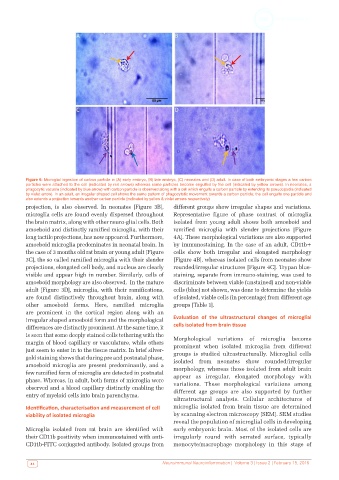
Figure 6: Microglial ingestion of carbon particle in (A) early embryo, (B) late embryo, (C) neonates and (D) adult. In case of both embryonic stages a few carbon
          particles were attached to the cell (indicated by red arrows) whereas some particles become engulfed by the cell (indicated by yellow arrows). In neonates, a
          phagocytic vacuole (indicated by blue arrow) with carbon particle is observed along with a cell which engulfs a carbon particle by extending its pseudopodia (indicated
          by violet arrow). In an adult, an irregular shaped cell shows the same pattern of phagocytotic movement towards a carbon particle; the cell engulfs one particle and
          also extends a projection towards another carbon particle (indicated by yellow & violet arrows respectively)
          projection, is also observed. In neonates [Figure 3B],   different groups show irregular shapes and variations.
          microglia cells are found evenly dispersed throughout   Representative figure of phase contrast  of microglia
          the brain matrix, along with other neuro-glial cells. Both   isolated from young adult shows both amoeboid and
          amoeboid and distinctly ramified microglia, with their   ramified microglia with slender projections [Figure
          long tactile projections, has now appeared. Furthermore,   4A]. These morphological variations are also supported
          amoeboid microglia predominates in neonatal brain. In   by immunostaining. In the case of an adult, CD11b+
          the case of 3 months old rat brain or young adult [Figure   cells  show  both  irregular  and  elongated  morphology
          3C], the so called ramified microglia with their slender   [Figure 4B], whereas isolated cells from neonates show
          projections, elongated cell body, and nucleus are clearly   rounded/irregular structures [Figure 4C]. Trypan blue-
          visible and appear high in number. Similarly, cells of   staining, separate from immuno-staining, was used to
          amoeboid morphology are also observed.  In the mature   discriminate between viable (unstained) and non-viable
          adult [Figure 3D], microglia, with their ramifications,   cells (blue) not shown, was done to determine the yields
          are found distinctively throughout brain, along with   of isolated, viable cells (in percentage) from different age
          other amoeboid forms. Here, ramified microglia      groups [Table 1].
          are prominent in the cortical region along with an
          irregular shaped amoeboid form and the morphological   Evaluation  of  the  ultrastructural  changes  of  microglial
          differences are distinctly prominent. At the same time, it   cells isolated from brain tissue
          is seen that some deeply stained cells tethering with the   Morphological variations of microglia become
          margin of blood capillary or vasculature, while others   prominent when isolated  microglia from  different
          just seem to enter in to the tissue matrix. In brief silver-
          gold staining shows that during pre and postnatal phase,   groups  is  studied  ultrastructurally.  Microglial  cells
                                                              isolated from neonates show rounded/irregular
          amoeboid microglia are present predominantly, and a   morphology, whereas those isolated from adult brain
          few ramified form of microglia are detected in postnatal   appear as  irregular,  elongated  morphology  with
          phase. Whereas, in adult, both forms of microglia were
          observed and a blood capillary distinctly enabling the   variations. These morphological variations among
                                                              different age groups are also supported by further
          entry of myeloid cells into brain parenchyma.
                                                              ultrastructural analysis. Cellular architectures of
          Identification, characterisation and measurement of cell   microglia isolated from brain tissue are determined
          viability of isolated microglia                     by scanning electron microscopy (SEM). SEM studies
                                                              reveal the population of microglial cells in developing
          Microglia isolated from rat brain are identified with   early embryonic brain. Most of the isolated cells are
          their CD11b positivity when immunostained with anti-  irregularly round with serrated surface, typically
          CD11b-FITC conjugated antibody. Isolated groups from   monocyte/macrophage morphology in this stage of


            44                                            Neuroimmunol Neuroinflammation |  Volume 3 | Issue 2 | February 15, 2016