
Page 52 - Read Online
P. 52
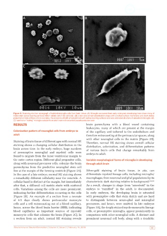
Figure 5: Scanning electron micrograph of isolated microglia cells from brain tissue. (A) Microglia recovered from early embryonic developing brain show many of
           them retain spines bearing surfaces which radiate out of the cell body ; (B) a few cells remain amoeboid in shape with a ruffled surface membrane and short stubby
           projections in late embryo; (C) in neonates, the presence of both amoeboid cells with spines bearing surfaces and also extended structure of amoeboid microglia cell;
           (D) ramified or “resting” microglia isolated from adult brain with extended pseudopodia are seen
           RESULTS                                             brain parenchyma with a blood vessel containing
                                                               leukocytes, many of which are present at the margin
           Colonization pattern of neuroglial cells from embryo to   of the capillary and tethered to the endothelium and
           adult                                               therefore extravaseting at the perivascular spaces, along
                                                               with other neuroglial cells in the matrix [Figure 2D].
           Staining of brain tissue of different ages with normal HE   Therefore, normal HE staining shows overall cellular
           staining shows a changing cellular distribution in the   distribution, colonization, and differentiation patterns
           brain across time. In the early embryo, huge numbers   of  various  brain  cells  that  change  remarkably  from
           of presumptive neuroglial and myeloid cells were    embryo to adult.
           found to migrate from the inner ventricular margin to
           the outer cortex region. Different glial progenitor cells,   Variable morphological forms of microglia in developing
           along with neuronal precursor cells, colonize the brain   through adult brain
           parenchyma from the predictive neuroglial stem cell
           line at the margin of the forming ventricle [Figure 2A].   Silver-gold  staining  of  brain  tissue,  in  situ,  can
           In the case of a late embryo, normal HE staining shows   differentiate myeloid lineage cells, including microglia/
           a remarkably different cellularity near the ventricle. A   macrophages, from neuronal and glial populations by its
           cellular band is distinct at the margin, but immediately   characteristic dark staining relative to background. [28,29]
           after that, a diffused cell matrix starts with scattered   As a result, changes in shape from “amoeboid” in the
           cells. Variations among the cells are more prominent,   embryo to “ramified” in the adult, is documented.
           indicating further differentiation occurring in the cells   In early embryos, the developing brain is saturated
           [Figure  2B].  An  example of  a section  from a  neonate   with presumptive cells that stain darkly and are hard
           of 4-5 days clearly shows perivascular monocyte     to distinguish between neuro-glial and microglial
           cells and a cell extravasating out of a blood capillary,   precursors, and hence, were omitted. In late embryos
           hence, across the blood brain barrier (BBB), indicating   [Figure 3A], the deeply stained myelo-monocytic lineage
           blood capillaries as important sources of myeloid/  cells, or presumptive microglia, appear prominently in
           monocytic cells that colonize the brain [Figure 2C]. In   comparison with other neuroglial cells. A distinct and
           a  section  from  an  adult,  normal  HE  staining  reveals   prominent neuronal cell body, along with a dendritic


           Neuroimmunol Neuroinflammation | Volume 3 | Issue 2 | February 15, 2016                           43