
Page 50 - Read Online
P. 50
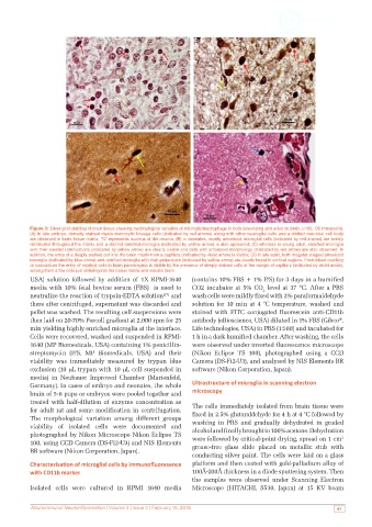
Figure 3: Silver gold staining of brain tissue showing morphological variation of microglia/macrophage in both developing and adult rat brain (×100, Oil immersion).
           (A) In late embryo, densely stained myelo-monocytic lineage cells (indicated by red arrows) along with other neuroglial cells and a distinct neuronal cell body
           are observed in brain tissue matrix. “N” represents nucleus of the neuron; (B) in neonates, mostly amoeboid microglial cells (indicated by red arrows) are evenly
           distributed throughout the matrix and a distinct ramified microglia (indicated by yellow arrow) is also appeared; (C) whereas in young adult, ramified microglia
           with their slender ramifications (indicated by yellow arrow) are clearly visible and cells with amoeboid morphology (indicated by red arrow) are also observed. In
           addition, the entry of a deeply stained cell into the brain matrix from a capillary (indicated by violet arrow) is visible; (D) in late adult, both irregular shaped amoeboid
           microglia (indicated by blue arrow) and ramified microglia with their projections (indicated by yellow arrow) are clearly found in cortical regions. From blood capillary
           or vasculature the entry of myeloid cells in brain parenchyma is visible by the presence of deeply stained cells at the margin of capillary (indicated by violet arrow),
           among them a few cells just entering into the tissue matrix and resides there
           USA) solution followed by addition of 1X RPMI-1640   (contains 10% FBS + 1% P/S) for 3 days in a humified
           media with 10% fetal bovine serum (FBS)  is used to   CO2 incubator at 5% CO  level at 37 ºC. After a PBS
                                                                                     2
           neutralize the reaction of trypsin-EDTA solution  and   wash cells were mildly fixed with 2% paraformaldehyde
                                                     [27]
           there after centrifuged, supernatant was discarded and   solution for 10 min at 4 °C temperature, washed and
           pellet was washed. The resulting cell suspensions were   stained with FITC conjugated fluorescein  anti-CD11b
           then laid on 20-70% Percoll gradient at 2,000 rpm for 25   antibody (eBioscience, USA) diluted in 1% FBS (Gibco ,
                                                                                                              ®
           min yielding highly enriched microglia at the interface.   Life technologies, USA) in PBS (1:500) and incubated for
           Cells were recovered, washed and suspended in RPMI-  1 h in a dark humified chamber. After washing, the cells
           1640 (MP Biomedicals, USA) containing 1% penicillin-  were observed under inverted fluorescence microscope
           streptomycin  (P/S, MP  Biomedicals,  USA)  and  their   (Nikon Eclipse TS 100), photographed using a CCD
           viability  was  immediately  measured  by  trypan  blue   Camera (DS-Fi2-U3), and analysed by NIS Elements BR
           exclusion (10 µL trypan with 10 µL cell suspended in   software (Nikon Corporation, Japan).
           media) in Neubauer Improved Chamber (Marienfeld,
           Germany). In cases of embryo and neonates, the whole   Ultrastructure of microglia in scanning electron
           brain of 5-6 pups or embryos were pooled together and   microscopy
           treated with half-dilution of enzyme concentration as
           for adult rat and some modification in centrifugation.   The cells immediately isolated from brain tissue were
           The morphological variation among different groups   fixed in 2.5% glutaraldehyde for 4 h at 4 °C followed by
           viability of isolated cells were documented and     washing in PBS and gradually dehydrated in graded
           photographed by Nikon Microscope Nikon Eclipse TS   alcohol and finally brought to 100% acetone. Dehydration
                                                                                                              2
           100, using CCD Camera (DS-Fi2-U3) and NIS Elements   were followed by critical-point drying, spread on 1 cm
                                                               grease-free glass slide placed on metallic stub with
           BR software (Nikon Corporation, Japan).
                                                               conducting silver paint. The cells were laid on a glass
           Characterisation of microglial cells by immunofluoresence   platform and then coated with gold-palladium alloy of
           with CD11b marker                                   100Å-200Å thickness in a diode sputtering system. Then
                                                               the samples were observed under Scanning Electron
           Isolated cells were cultured in RPMI 1640 media     Microscope (HITACHI, S530, Japan) at 15 KV beam


           Neuroimmunol Neuroinflammation | Volume 3 | Issue 2 | February 15, 2016                           41