
Page 28 - Read Online
P. 28
metastases tend to have a thicker ring enhancement and a
reduced diffusion in the necrotic center. Brain metastases
frequently have a thicker ring enhancement, but typically
have no reduced diffusion in the necrotic center. [5]
Definitive diagnosis is established either by isolation of
the fungus from a culture or direct visualization on the
histological slides. Isolation from the CSF is uncommon.
In a case series of 22 patients with CNS blastomycosis, CSF
[2]
cultures were positive only in 2 patients. Serologic testing
is generally considered not to be useful in blastomycosis
due to high cross-reactivity with other endemic mycoses.
Antigen testing may be positive in the urine and serum.
PCR is rarely used and typically not commercially
available.
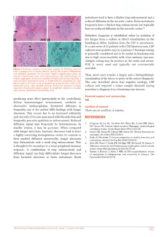
Figure 3: Histologic analysis of the biopsy sample. (a) Histologic sections
show a background of reactive fibrosis (arrow) with nodular inflammatory
cell infiltrates (between vertical lines). Higher magnification (×10); (b) Thus, most cases require a biopsy and a histopathologic
chronic inflammatory cells: small mononuclear cells (arrow head) and
nodular aggregates of pale pink “epithelioid” histiocytes (arrow) imparting a examination of the tissue to arrive at the correct diagnosis.
granulomatous appearance (×20); (c) Small spherical organisms are seen The case described above had negative serology, CSF
(arrow head) and focally a few neutrophils are present in the center of the
granuloma (arrow, ×40); (d) GMS staining confirms the presence of fungal culture and required a tissue sample obtained during
organisms that morphologically appear as small dark stained round yeast resection to diagnose it as a blastomycosis abscess.
forms (arrow). Blastomyces dermatitidis (×40).
Financial support and sponsorship
producing mass effect (particularly in the cerebellum), Nil.
diffuse leptomeningeal enhancement, cerebritis or
obstructive hydrocephalus. Restricted diffusion is Conflicts of interest
frequently one of the earliest MRI findings with fungal There are no conflicts of interest.
abscesses. This occurs due to an increased cellularity
and viscosity of the pus associated with the infection and REFERENCES
frequently precedes gadolinium enhancement. Reduced
diffusion signal may frequently be heterogeneous. In 1. Chapman W, Lin AC, Hendricks KA, Nolan RL, Currier MM, Morris
smaller lesions, it may be punctate. When compared KR, Turner HR. Endemic blastomycosis in Mississippi: epidemiological
and clinical studies. Semin Respir Infect 1997;12:219-28.
with fungal infections, bacterial abscesses tend to have 2. Kravitz GR, Davies SF, Eckman MR, Sarosi GA. Chronic blastomycotic
a highly restricting homogeneous center. In contrast to meningitis. Am J Med 1981;71:501-5.
their marked diffusion abnormality, fungal abscesses 3. Szabo G, Mandrekar P. A recent perspective on alcohol, immunity, and
host defense. Alcohol Clin Exp Res 2009;33:220-32.
may demonstrate only a weak ring enhancement. This 4. Bush JW, Wuerz T, Embil JM, Del Bigio MR, McDonald PJ, Krawitz S.
is thought to be secondary to a weak peripheral immune Outcomes of persons with blastomycosis involving the central nervous
response. A combination of ring enhancement and system. Diagn Microbiol Infect Dis 2013;76:175-81.
diffusion signal can help differentiate fungal abscesses 5. Starkey J, Moritani T, Kirby P. MRI of CNS fungal infections: review
of aspergillosis to histoplasmosis and everything in between. Clin
from bacterial abscesses or brain metastases. Brain Neuroradiol 2014;24:217-30.
Neuroimmunol Neuroinfammation | Volume 3 | Issue 1 | January 20, 2016 19