
Page 27 - Read Online
P. 27
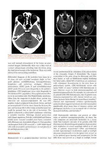
Figure 1: Axial head computed tomography image revealing a predominantly
          hypodense mass lesion in the cerebellum, centered at the vermis. The
          lesion compresses the fourth ventricle (arrow pointing to the mass)


          was avid internal enhancement of the lesion on post-  Figure  2: Axial magnetic resonance imaging images of fluid-attenuated
          contrast images. Additionally there was a thin tract of   inversion recovery (a) and T1 postcontrast (b) at initial presentation and then
                                                              postoperatively (c and d) revealing complete radiographic resection of the lesion
          contrast enhancement extending from the lesion along
          the right lateral margin of the brainstem. There was mild
          edema of the surrounding cerebellum.                occurs predominantly by inhalation of the microconidia
                                                              of the dimorphic fungus  B.  dermatitidis. The fungus
          Differential  diagnosis  of  the  posterior  fossa  mass  in  a   is prevalent in the areas along the Mississipi and Ohio
          41  years  old  male  included  neoplastic  (high-  or  low-  river basins as well as Midwestern regions bordering
          grade gliomas, medulloblastoma, hemangioblastoma,   the Great Lakes. About 91% of infections are pulmonary
          lymphoma    and   infectious  etiologies.  Pilocytic  with subsequent dissemination to the skin, bone or
          astrocytomas (WHO grade I) and medulloblastomas     genitourinary system. CNS involvement occurs only
                                                                                 [1]
          (WHO grade IV) occur more frequently in the pediatric   rarely 5-10% of cases.  Isolated CNS blastomycosis is
                                                              rare. Infection occurs in both immunocompetent and
          population. CNS lymphomas occur more frequently in   immunosuppressed hosts. For example, in a case series of
          the elderly or HIV+ population. Hemangioblastomas often   22 patients with CNS blastomycosis, only 12 patients were
          occur in association with Von Hippel-Lindau syndrome.   immunocompromised (i.e. HIV, chronic steroid use, anti-
          Other than the suspicion of alcohol abuse, patient was   tumor necrosis factor therapy for more than 6 months).
                                                                                                             [2]
          otherwise not immunocompromised. He was HIV         Clinical and experimental evidence (predominantly
          negative, lacked peripheral leukocytosis, fevers, and had   animal studies) suggests that chronic alcohol consumption
          an unremarkable CT of the chest. Given the location, the   significantly  alters  many  lines  of  immune  system  and
          size, and the mass effect of the lesion, no lumbar puncture   predisposes alcoholics to an increased risk of infection,
          could be pursued due to the risk of herniation. The   increased morbidity, and mortality.
                                                                                            [3]
          patient underwent a suboccipital craniotomy achieving
          a complete resection. Pathology showed granulomas   CNS blastomycotic infection can present as either
          with fungal organisms. Periodic acid-Schieff and Giemsa   acute or indolent meningoencephalitis. At times, the
          (GMS)  stains  revealed  rounded  yeast  forms  consistent   only symptoms are intractable headaches. In a recent
          with Blastomycosis dermatitidis [Figure 3]. Post-operative   study that evaluated outcomes of 16 patients with CNS
          cerebrospinal fluid (CSF) revealed > 1,000 White blood   blastomycosis, the most frequent symptoms at presentation
          cells with neutrophilic predominance, normal glucose at   were headaches or a focal neurologic deficit in 63 and
                                                                                         [4]
          64 and high protein at 300.                         56% of patients respectively.  Other manifestations
                                                              include leptomeningeal involvement, single or multiple
          DISCUSSION                                          abscesses intracranially or in the spinal cord, as well as
                                                              in the epidural space causing cord compression. On the
          Blastomycosis is a pyogranulomatous infection that   MRI  these  may present  as  a  single  or  multiple  lesions


            18                                             Neuroimmunol Neuroinfammation | Volume 3 | Issue 1 | January 20, 2016