
Page 25 - Read Online
P. 25
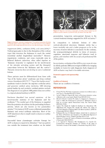
Figure 5: Magnetic resonance angiography showing patent basilar tip and
                                                              posterior cerebral arteries. (a) Anterior view; (b) lateral view right side

                                                              presentation. Long‑term anticoagulant therapy is the
                                                              current treatment strategy suggested for AOP occlusion. [7]


          Figure 4: Magnetic resonance imaging brain T2‑weighted sequences showing   In  comparison  to  ischemic  lesions  of  other
          V‑shaped hyper‑intense signal areas in the pial surface of the midbrain
          adjacent to the interpeduncular fossa without any abnormal signs in this region  cortical‑subcortical structures, thalamic stroke has a
                                                              lower mortality rate and a better prognosis as far as the
                                                         [2]
          impairment (58%), confusion (53%), and coma (42%).    recovery of motor deficits is concerned. On the contrary,
          Vertical gaze palsy is due to the disruption of the cortical   the neuropsychological deficits in terms of memory,
          input that traverses the thalamus to reach the rostral   cognition, emotional response and behavior tend to
          interstitial medial longitudinal fasciculus.  Memory   persist, and interfere with the social and professional life
                                                [3]
          impairment,  confusion,  and  coma  classically  seen  in   of the patient.
          bilateral thalamic infarction, often called together as
          “thalamic dementia” is explained by the involvement   In conclusion, occlusion of the AOP is a rare cause of coma
          of the reticular activating system and the disrupted   in elderly patients. Diffusion‑weighted MRI is the imaging
                                                         [4]
          connections between the thalamus and the cortex.    modality of choice for early diagnosis. Early diagnosis of
          Thalamic dementia does not develop in case of unilateral   AOP occlusion may lead to favorable outcomes.
          lesions.
                                                              Financial support and sponsorship
          These patients must be differentiated from those with   Nil.
          “top of the basilar artery” syndrome and deep cerebral
          venous thrombosis (DCVT). [5,6]  “Top of the basilar artery”   Conflicts of interest
          syndrome tends to involve the superior cerebellar artery   There are no conflicts of interest.
          and posterior cerebral artery territories. MRI showing
          patent basilar tip and posterior cerebral arteries exclude   REFERENCES
          this diagnosis in our patient. MRI pattern does not confine
          to a typical arterial territory in DCVT.            1.   Chang YM, Fan YK. Artery of percheron occlusion in an elderly male: a
                                                                 case report. J Clin Med Res 2015;7:126‑8.
                                                              2.   Lamot U, Ribaric I, Popovic KS. Artery of Percheron infarction: review
          Percheron described four normal variations  of the     of literature with a case report. Radiol Oncol 2015;49:141‑6.
          neurovascular anatomy of the thalamus and the       3.   Monet P, Monet P, Garcia PY, Saliou G, Spagnolo S, Desblache J,
                                                                 Franc J, Vallée JN, Deramond H, Lehmann P. Bithalamic infarct: is
          midbrain.  The medial part of the thalamus is supplied   there  an  evocative  aspect?  Radioclinical  study.  Rev  Neurol  (Paris)
                  [2]
          from the posterior circulation via the perforating thalamic   2009;165:178‑84.
          arteries,  which are also  known as  the paramedian   4.   Lazzaro NA, Wright B, Castillo M, Fischbein NJ, Glastonbury CM,
                                                                 Hildenbrand PG, Wiggins RH, Quigley EP, Osborn AG. Artery of
          arteries.  Occlusion of AOP causes a bilateral paramedian   percheron infarction: imaging patterns and clinical spectrum. AJNR Am
                [2]
          thalamic  and  rostral  midbrain  infarction.  Most  of the   J Neuroradiol 2010;31:1283‑9.
          AOP infarction is due to small vessel occlusion or cardiac   5.   Rodriguez EG, Lee JA. Bilateral thalamic infarcts due to occlusion of
                                                                 the Artery of Percheron and discussion of the differential diagnosis of
          embolism. [5]                                          bilateral thalamic lesions. J Radiol Case Rep 2013;7:7‑14.
                                                              6.   Kostanian V, Cramer SC. Artery of Percheron thrombolysis. AJNR Am
          Successful tissue plasminogen activator therapy for    J Neuroradiol 2007;28:870‑1.
                                           [7]
          AOP occlusion is reported in literature,  but our patient   7.   Li X, Agarwal N, Hansberry DR, Prestigiacomo CJ, Gandhi CD.
                                                                 Contemporary therapeutic strategies for occlusion of the artery of
          was outside the treatment time window on the initial   Percheron: a review of the literature. J Neurointerv Surg 2015;7:95‑8.


            16                                             Neuroimmunol Neuroinfammation | Volume 3 | Issue 1 | January 20, 2016