
Page 30 - Read Online
P. 30
the capacity of stem cells to self-renew and generate THE SVZ IN THE ADULT HUMAN BRAIN
differentiating cells while also maintaining their pool, it
has been proposed that SVZ stem cells could play a role The identification of neurogenic niches in rodents [4]
in tumorigenesis. This hypothesis has been supported has challenged the long-standing notion that the
by studies using genetically-engineered animal models mammalian brain was a quiescent organ characterized
[5]
where the key genetic alterations of HGG occur only in by lack of neurogenesis postnatally. In the adult
neural stem/precursor cells of the SVZ. mammalian brain, neurogenesis occurs in 2 germinal
[6]
regions: the SVZ and the subgranular layer (SGL) of
[7]
The advent of high-resolution genomic techniques the dentate gyrus of the hippocampus. Several works
gave us the unique opportunity to overcome the on the cellular organisation of the SVZ in rodents have
challenges associated with studies in the human brain revealed the existence of neural stem cells that express
the astrocytic marker glial fibrillary acidic protein
of HGG where only a small amount of tumor tissue (GFAP) and give rise to neurons. When compared to
is available and longitudinal studies to assess tumor the SGL, the SVZ represents the most abundant source
development are not possible. We developed a real-time of neurons. [8-11] More recently, studies on the adult
fluorescence-guidedtiple-sampling (FGMS) strategy human brain have shown that the SVZ retains the
based on 5-aminolevulinic acid to identify cancer same functional properties of the rodent brain, but the
stem cells in different tumor regions and we used GFAP+ve cells are organised in a ribbon. [12,13] However,
[1]
this approach to describe the extent of spatial genetic important differences exist between the human and
intra- tumor heterogeneity in HGG [1,2] and to reconstruct rodent SVZ: (1) in humans, the SVZ is positioned in the
tumorigenesis. In parallel, we derived cancer stem cells wall of the lateral ventricles and is characterized by 4
[2]
from the tumor mass and the SVZ of the same patients layers. SVZ astrocytes are organised in ribbons separated
and we showed that drug-resistant cells are present in from the ependymal layer by a hypocellular gap, that is
this niche. These findings have implications for the a reminiscence of the neuronal formation and migration
[3]
[14]
development of new therapeutic approaches targeting occurring at embryonic stages [Figure 1]. Interestingly,
the SVZ. the terms SVZ and SEZ have been used interchangeably,
however they describe specifically these layers with the
inclusion or not of the ependymal layer [Figure 1]; (2)
the number of actively proliferating cells in human SVZ
is very low in comparison to rodents; [12,15] and (3) the
evidence of the existence of neural stem cells in vivo is
still missing in humans, whereas it is well established
in rodents.
Accumulating evidence points out to the influence of
pathological conditions on neurogenesis. These include
infections, inflammations, stroke, epilepsy, tumors
and neurodegenerative disorders. [16,17] For instance, in
Huntington’s disease an increase in cell proliferation
and neurogenesis occur in the SVZ of disease brains.
[18]
Extending our understanding of the biology of the
human SVZ might lead to the identification of novel
therapeutic interventions against the large spectrum of
diseases affecting the brain.
THE SVZ AS INFLAMMATORY RESERVOIR
In HGG, the onset of malignant transformation can be
seen as a traumatic event that can initiate inflammation.
This can then persist during the subsequent phases
of tumor growth: promotion and progression.
[19]
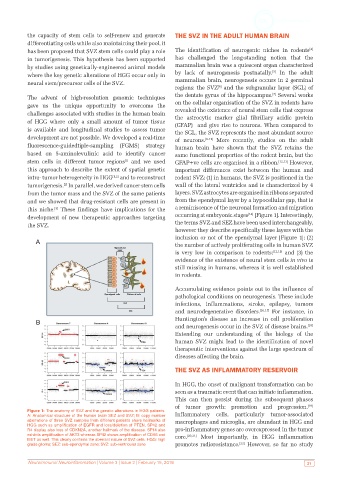
Figure 1: The anatomy of SVZ and the genetic alterations in HGG patients.
A: Anatomical structure of the human brain SEZ and SVZ; B: copy number Inflammatory cells, particularly tumor-associated
aberrations of three SVZ samples from different patients share hallmarks of macrophages and microglia, are abundant in HGG and
HGG such as amplification of EGFR and loss/deletion of PTEN. SP42 and
R4 display also loss of CDKN2A, another hallmark of the disease. SP14 also pro-inflammatory genes are overexpressed in the tumor
exhibits amplification of AKT3 whereas SP42 shows amplification of CDK6 and core. [20,21] Most importantly, in HGG inflammation
MET as well. This clearly confirms the aberrant nature of SVZ cells. HGG: high
[22]
grade glioma; SEZ: sub-ependymal zone; SVZ: sub-ventricular zone promotes radioresistance. However, so far no study
Neuroimmunol Neuroinflammation | Volume 3 | Issue 2 | February 15, 2016 21