
Page 32 - Read Online
P. 32
responsible for tumor re-initiation following chemo- can be further investigated to elucidate the process of
therapy. In support of these findings it was also noted oncogenesis in HGG patients. The limited availability
[54]
that p53 mutations preferentially occur in the SVZ. [55] of tissue samples and the clinical complex scenario
at the time of surgery make it difficult to reconstruct
Collectively, these results raise the question on whether the initial steps of tumor development and alternative
cancer stem cells directly derive from SVZ stem cells. methods are needed. Given the critical functional
Although mouse model studies have indicated that this role of the SVZ in the adult human brain, it has been
is the case, these findings have been severely hampered speculated that this niche might play a role in neuro-
by a limited representation of the aberrant genetic oncogenesis. This has been the focus of our recent
landscape of HGG and the use of markers that poorly study on HGG patients. [3]
discriminate between stem cells and precursor cells. [32]
More recently, the same question has been addressed by THE SVZ AS A SOURCE OF TUMOR CELLS IN
using a transgenic cell-labelling system known as mosaic HGG PATIENTS
analysis with double markers. Using this model, it
[56]
has been proposed that the cells of origin in HGG are The identification of cancer stem cells from human
oligodendrocyte precursor cells, thus challenging the HGG has represented a novel tool to develop therapeutic
notion that HGG may originate from transformation and strategies [57,58] and these cells have been proposed
expansion of the neural stem cell pool. as a model that more closely represents the human
disease. We took advantage of these findings to
[59]
Although the debate about the cell of origin in HGG objectively interrogate primary HGG in humans using
is still open, the above studies have helped define the a neurosurgical techniques based on FGMS. In the
potential targets of malignant transformation that clinic fluorescence-guided resection has resulted in
enhanced cytoreduction and improved progression-free
[60]
survival in patients in a randomized Phase III trial.
We have adapted this technology to allow the objective
identification of tumor tissue based on combining
fluorescence emission and neuroanatomical landmarks
and we have recently demonstrated that this technique
can be successfully employed to characterize cancer
stem cells derived from fluorescent and non-fluorescent
material in HGG patients. [1]
Quite unexpectedly, we observed for the first time
that fluorescent material is present in the SVZ of 42
out of 65 HGG patients who underwent surgery using
fluorescence-guided resection and we isolated tissue
from the tumor mass and the SVZ. Using these samples
we reported that the SVZ contains malignant cells
that contribute to tumor growth. This has never been
[3]
demonstrated in humans, but similar observations have
been reported in mouse models of HGG. [46,53-55,61]
Importantly, the phylogenetic relationship between
SVZ and tumor in these patients identifies the SVZ as
a reservoir of tumor cells (either early tumor clones or
late-emergent clones that develop during HGG growth)
that need to be therapeutically targeted. Thus, we
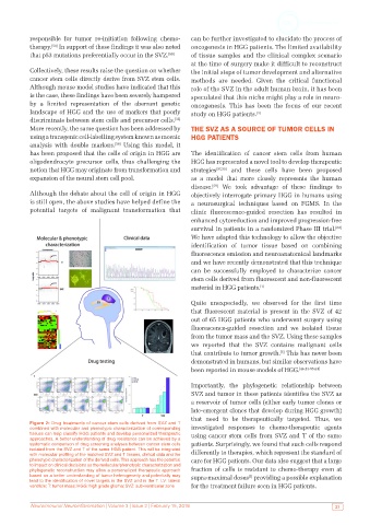
Figure 2: Drug treatments of cancer stem cells derived from SVZ and T
combined with molecular and phenotypic characterization of corresponding investigated responses to chemo-therapeutic agents
tissues can help classify HGG patients and develop personalized therapeutic using cancer stem cells from SVZ and T of the same
approaches. A better understanding of drug resistance can be achieved by a
systematic comparison of drug screening analyses between cancer stem cells patients. Surprisingly, we found that such cells respond
isolated from the SVZ and T of the same HGG patient. This will be integrated differently to therapies, which represent the standard of
with molecular profiling of the matched SVZ and T tissues, clinical data and the
phenotypic characterization of the derived cells. This approach has the potential care for HGG patients. Our data also suggest that a large
to impact on clinical decisions as the molecular/phenotypic characterization and
phylogenetic reconstruction may allow a personalized therapeutic approach fraction of cells is resistant to chemo-therapy even at
based on a better understanding of tumor heterogeneity and potentially may supra-maximal doses providing a possible explanation
[3]
lead to the identification of novel targets in the SVZ and in the T. LV: lateral
ventricle; T: tumor mass; HGG: high grade glioma; SVZ: sub-ventricular zone for the treatment failure seen in HGG patients.
Neuroimmunol Neuroinflammation | Volume 3 | Issue 2 | February 15, 2016 23