
Page 37 - Read Online
P. 37
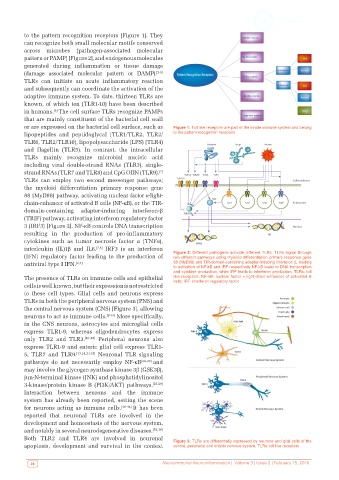
to the pattern recognition receptors [Figure 1]. They
          can recognize both small molecular motifs conserved
          across microbes (pathogen-associated molecular
          pattern or PAMP) [Figure 2], and endogenous molecules
          generated during inflammation or tissue damage
          (damage associated molecular pattern or DAMP). [2-5]
          TLRs can initiate an acute inflammatory reaction
          and subsequently can coordinate the activation of the
          adaptive immune system. To date, thirteen TLRs are
          known, of which ten (TLR1-10) have been described
                    [6]
          in humans. The cell surface TLRs recognize PAMPs
          that are mainly constituent of the bacterial cell wall
          or are expressed on the bacterial cell surface, such as   Figure 1: Toll like receptors are part of the innate immune system and belong
          lipopeptides and peptidoglycal (TLR1/TLR2, TLR2/    to the pattern recognition receptors
          TLR6, TLR2/TLR10), lipopolysaccharide (LPS) (TLR4)
          and flagellin (TLR5). In contrast, the intracellular
          TLRs mainly recognize microbial nucleic acid
          including viral double-strand RNAs (TLR3), single-
          strand RNAs (TLR7 and TLR8) and CpG ODN (TLR9). [7]
          TLRs can employ two second messenger pathways;
          the  myeloid differentiation primary response  gene
          88 (MyD88) pathway, activating nuclear factor κ-light-
          chain-enhancer of activated B cells (NF-κB), or the TIR-
          domain-containing adapter-inducing  interferon-β
          (TRIF) pathway, activating interferon regulatory factor
          3 (IRF3) [Figure 2]. NF-κB controls DNA transcription
          resulting in the production of pro-inflammatory
          cytokines such as  tumor necrosis factor  α  (TNFα),
          interleukin (IL)1β and IL6. [7,8]  IRF3 is an interferon
          (IFN)  regulatory factor leading to  the production  of   Figure 2: Different pathogens activate different TLRs. TLRs signal through
                                                              two different pathways using myeloid differentiation primary response gene
          antiviral type I IFN. [2,7]                         88 (MyD88) and TIR-domain-containing adapter-inducing interferon β, leading
                                                              to activation of NF-kB and IRF respectively NF-kB leads to DNA transcription
                                                              and cytokine production, while IRF leads to interferon production. TLRs: toll
          The presence of TLRs on immune cells and epithelial   like receptors; NF-kB: nuclear factor κ-light-chain-enhancer of activated B
                                                              cells; IRF: interferon regulatory factor
          cells is well known, but their expression is not restricted
          to these cell types. Glial cells and neurons express
          TLRs in both the peripheral nervous system (PNS) and
          the central nervous system (CNS) [Figure 3], allowing
          neurons to act as immune cells. [9-15]  More specifically,
          in the CNS neurons, astrocytes and microglial cells
          express TLR1-9, whereas oligodendrocytes express
          only TLR2 and TLR3.  [16-20]  Peripheral neurons also
          express TLR1-9 and enteric glial cell express TLR1-
          5, TLR7 and TLR9. [13,14,21-23]  Neuronal TLR signaling
          pathways do not necessarily employ NF-κB [24-26]  and
          may involve the glycogen synthase kinase 3β (GSK3β),
          jun-N-terminal kinase (JNK) and phosphatidylinositol
          3-kinase/protein  kinase B (PI3K/AKT) pathways. [27-29]
          Interaction between neurons and the immune
          system has already been reported, setting the scene
          for neurons acting as immune cells. [30-34]  It has been
          reported that neuronal TLRs are involved in the
          development and homeostasis of the nervous system,
          and notably in several neurodegenerative diseases. [35,36]
          Both TLR2 and TLR4 are involved in neuronal         Figure 3: TLRs are differentially expressed by neurons and glial cells of the
          apoptosis, development and survival in the context   central, peripheral and enteric nervous system. TLRs: toll like receptors


            28                                            Neuroimmunol Neuroinflammation |  Volume 3 | Issue 2 | February 15, 2016