
Page 104 - Read Online
P. 104
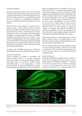
minimal CSF findings. [13]                          indirect immunofluorescence on a substrate of mouse or rat
                                                               brain sections. TBA is an excellent screening method, as the
           The electroencephalogram (EEG) is almost always abnormal   antibody target antigen can be suspected from the staining
           in all types of AIE, showing focal or diffuse slow activity that   pattern (e.g. neuropil), although it must be confirmed by more
           can be associated with focal or multifocal epileptic discharges.   specific  techniques  [Figure  2A].  As  regards  the  detection
           Except for a pattern referred to as extreme delta brush, that   of onconeural antibodies (e.g. Hu, Ma2, Ri, amphiphysin),
                                                         [14]
           may  occur  in  patients  with  anti-NMDAR  encephalitis,    commercial immunoblots with recombinant proteins for
           there are no pathognomonic EEG abnormalities for any AIE   the most common autoantibodies are widely available.
           subtypes.                                           On the other hand, the gold standard for neuronal surface
                                                               autoantibody detection is CBA, in which cells (e.g. human
           MRI of the brain is often diagnostic in patients with LE,   embryonic kidney 293 cells) expressing the appropriate
           usually  showing  increased  Fluid  Attenuated  Inversion   antigens  are  incubated with  patients’  serum/CSF,  and
           Recovery/T2-weighted  (FLAIR/T2)  signal involving  one  or   antibodies are identified by indirect immunofluorescence
           both temporal lobes, without contrast enhancement. Similar   [Figure 2C]. This is a highly sensitive technique, but it is time
           findings can, however, occur in patients with herpes simplex   consuming and requires specific facilities and expertise.
           encephalitis or medial temporal lobe seizures. In NMDAR
           encephalitis  brain  MRI  is  normal  in  up  to  66%  of  cases,   Indirect immunofluorescence on live hippocampal or cortical
           while the remaining patients may have unspecific cortical   murine neurons is used as a screening method, in some
           or subcortical FLAIR/T2 abnormalities, sometimes involving   laboratories, for the detection of antibodies binding neuronal
           the posterior fossa or medial temporal regions, often with   plasma membrane proteins [Figure 2B].
           small areas of demyelination, and more rarely with extensive
           demyelinating abnormalities. [15]                   The most reliable method of detecting antibodies specific
                                                               for neuronal plasma membrane antigens involves using
           In patients with GABA(A)R antibodies, brain MRI often   a combination of TBA and indirect immunofluorescence
           shows multifocal cortical-subcortical FLAIR abnormalities. [16]  on live neuronal cultures as screening following by
                                                               confirmatory CBA.
           Detection of autoantibodies
           Several techniques are available for intracellular and   Caveats in the diagnosis
           synaptic antibody detection, for example, tissue-based   Standardized methods of antibody testing are critical to
           assays (TBA; in-house or commercially available), cell-based   ensure the correct diagnosis of AIE. However there are few
           assay (CBA; in-house or commercially available), indirect   studies, mainly in anti-NMDAR encephalitis, [17-19]  comparing
           immunofluorescence on live hippocampal or cortical   the sensitivity and specificity of different techniques
           neurons (in-house) and immunoprecipitation (IP; in-house).   in serum and CSF samples from patients with AIE. For
                                                                                        [20]
           In TBA, antibodies in patient serum or CSF are detected by   example, Gresa-Arribas et al.  examined paired serum


           Figure 2: IgG in the CSF from a patient with anti-NMDAR encephalitis bind to the neuropil of the mouse hippocampus (A), to the cell-surface of live, non-
           permeabilized mouse hippocampal neurons (B) and to the plasma membrane of HEK293 cells expressing NMDAR (C). anti-NMDAR: anti-N-methyl-d-aspartate
           receptor


           Neuroimmunol Neuroinflammation | Volume 3 | April 19, 2016                                       95