
Page 103 - Read Online
P. 103
the search for the presence of an underlying neoplasm [Figure
1].
Clinical presentation
AIE is usually a multistage process. Most of these disorders
have a rapid course, developing over a few days or weeks,
with behavioral and memory alteration, decreased level of
consciousness and seizures. This clinical picture is typical
of limbic encephalitis (LE). However, the severity and
predominance of some symptoms over others may help the
clinician in the diagnosis of different AIE subtypes and may
lead the search for specific antibodies [Table 1]. For example,
both GABA(B)R and gamma-aminobutyric acid A [GABA(A)
R] antibodies are typically associated with refractory
seizures, patients with leucine-rich glioma-inactivated
[4,5]
1 (LGI1) autoantibodies can present with facio-brachial
dystonic seizuresand hyponatremia caused by syndrome of
[6]
inappropriate antidiuresis (SIAD), while AMPAR-antibodies
[7]
are frequently found in patients with LE or psychosis. In
anti-NMDAR encephalitis, psychiatric disturbances are the
[8]
most frequent symptoms of onset in women, while seizures
are prominent in men. [9]
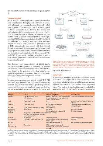
Figure 1: Flowchart summarizing a preferred diagnostic approach to AIE. AIE:
Anti-neuronal autoimmune encephalitis; CSF: cerebrospinal fluid; CBA: cell-
The detection and characterization of IgLON family based assay; EEG: electroencephalogram; TBA: tissue-based assays; CT:
member 5 antibodies represents an interesting link between computed tomography; PET: positron emission tomography; MRI: magnetic
resonance imaging; IVIG: intravenous immunoglobulin; PLEX: plasmapheresis
autoimmunity and neurodegeneration. These autoantibodies
were found to be associated with sleep disturbances, psychiatric dysfunction. [11]
cognitive impairment, the movement disorder and brainstem
symptoms with a chronic progressive course. [10] Ancillary tests
At presentation, about 80% of patients with AIE have a mild-
In some cases, symptoms may extend beyond CNS: AIE to-moderate CSF lymphocytic pleocytosis (usually < 100
associated with autoantibodies to dipeptidyl-peptidase-like white blood cells/L), 30% have a mild-to-moderate increase
protein-6 may present with diarrhea poorly responsive to inprotein concentration, and 50-60% have oligoclonal
symptomatic treatment and significant weight loss that can bands. In contrast to most autoimmune encephalitides,
[12]
precede neurological symptoms including brainstem and encephalitis with LGI1-IgGusually occurs with normal or
Table 1: Neuronal surface autoantibodies, associated tumors and clinical syndromes
Antigen Tumor Clinical symptoms Clinical clues
NMDAR Ovarian teratoma (58%) Memory impairment, psychosis (mainly Orobuccal dyskinesia; dysautonomia
< 18 years old in women), seizures (mainly in men),
central hypoventilation
LGI1 Thymoma (< 10%) LE Hyponatremia; faciobrachial dystonic
seizures
CASPR2 Thymoma (38%) Encephalitis/Morvansynd/ Peripheral nerve hyperexcitability;
neuromyotonia neuropathic pain
AMPAR SCLC, breast, thymoma (60-70%) LE, psychosis
GABA(B) R SCLC (50%) LE, ataxia Refractory seizures
GABA(A) R - Status epilepticus, seizures, LE Refractory seizures
mGluR1 Hodgkin and non Hodgkin lymphoma Cerebell arataxia
(e.g. cutaneus lymphoma); prostate
adenocarcinoma [3]
mGluR5 M. Hodgkin Ophelia syndrome Memory impairment
DPPX (Kv4.1) Follicular B cell, lymphoma, CLL Hallucinations, agitation, myoclonus, Diarrhea
tremor, SPS
IgLON5 - Brain stem dysfunction, LE Non-REM and REM-sleep disorder
GlyR Thymoma SPS, progressive encephalitis
Dopamine 2R - Basal ganglia encephalitis, Sydenham
Chorea
NMDAR: N-methyl-d-aspartate receptor; LGI1: leucine-rich glioma-inactivated 1; CASPR2:contactin-associated protein-like 2; AMPAR: amino-3-hydroxy-5-hydroxy-5-methyl-
4-isoxazolepropionic acid receptor; GABA A/B R: gamma-aminobutyric acid A/B receptor; mGluR1/5: metabotropic glutamate receptor type 1/5; DPPX: dipeptidyl-peptidase-
like protein-6; GlyR: Glycine receptor; CLL: chronic lymphatic leukemia; SCLC: small cell lung cancer; LE: limbic encephalitis; SPS: stiff-person syndrome; IgLON5: IgLON family
member 5
94 Neuroimmunol Neuroinflammation | Volume 3 | April 19, 2016