
Page 163 - Read Online
P. 163
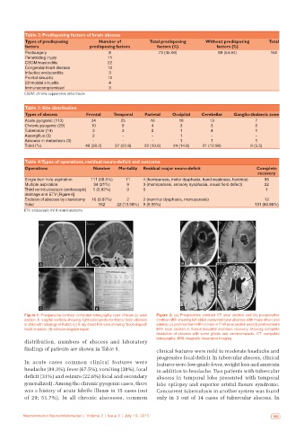
Table 2: Predisposing factors of brain abscess
           Types of predisposing        Number of           Total predisposing   Without predisposing     Total
           factors                  predisposing factors      factors (%)             factors (%)
           Postsurgery                      8                  73 (45.06)             89 (54.94)           162
           Penetrating injury              11
           CSOM/mastoiditis                22
           Congenital heart disease        10
           Infective endocarditis           3
           Frontal sinusitis               12
           Ethmoidal sinusitis              4
           Immunocompromised                3
           CSOM: chronic suppurative otitis media


           Table 3: Site distribution
           Types of abscess        Frontal   Temporal     Parietal   Occipital  Cerebellar   Ganglio‑thalamic zone
           Acute pyogenic (113)      34         25          16         18          13                7
           Chronic pyogenic (29)     10          9          4           3           3                0
           Tubercular (14)           3           3          2           1           4                1
           Aspergillus (3)           2           ‑           ‑          1           ‑                ‑
           Abscess in metastesis (3)  ‑          ‑           ‑          1           1                1
           Total (%)              49 (30.2)   37 (22.8)   22 (13.6)  24 (14.8)  21 (12.96)         9 (5.5)


           Table 4: Types of operations, residual neuro‑deficit and outcome
           Operations                 Number   Mortality  Residual major neuro‑deficit               Complete
                                                                                                      recovery
           Single burr hole aspiration  111 (68.5%)  11  4 (hemiparesis, motor dysphasia, hand weakness, footdrop)  96
           Multiple aspiration        34 (21%)     9     3 (monoparesis, sensory dysphasia, visual field defect)  22
           Third ventriculoscopic (endoscopic)   1 (0.62%)  0  0                                         1
           drainage and ETV [Figure 6]
           Excision of abscess by craniotomy  16 (9.87%)  2  2 (nominal dysphasia, monoparesis)          12
           Total                        162    22 (13.58%)  9 (5.55%)                                131 (80.86%)
           ETV: endoscopic third ventriculostomy


                                                                                       b
                 a                 b                                    a


                 c                     d                                c             d
           Figure 1: Preoperative contrast computed tomography scan of brain (a: axial   Figure  2:  (a) Preoperative contrast CT axial section and  (b) preoperative
           section; b: sagittal section) showing right‑sided posterior frontal brain abscess   contrast MRI showing left sided paraventricular abscess with mass effect and
           in child with tetralogy of Fallot; (c) X‑ray chest P/A view showing “boot shaped”   edema; (c) posttreatment MRI of brain in T1W axial section and (d) posttreatment
           heart shadow; (d) echocardiogram report            MRI axial section in fluid‑attenuated inversion recovery showing complete
                                                              resolution of abscess with some gliosis and cerebromalacia. CT: computed
                                                              tomography; MRI: magnetic resonance imaging
           distribution, numbers of abscess and laboratory
           findings of patients are shown in Table 5.         clinical features were mild to moderate headache and

                                                              progressive focal deficit. In tubercular abscess, clinical
           In  acute cases common clinical features were      features were low-grade fever, weight loss and anorexia
           headache (89.3%), fever (67.5%), vomiting (38%), focal   in addition to headache. Two patients with tubercular
           deficit (31%) and seizure (22.6%) focal and secondary   abscess in temporal lobe presented with temporal
           generalized). Among the chronic pyogenic cases, there   lobe epilepsy and superior orbital fissure syndrome.
           was a history of acute febrile illness in 15 cases (out   Concurrent tuberculosis in another system was found
           of 29; 51.7%). In all chronic abscesses, common    only in 3 out of 14 cases of tubercular abscess. In


   154  Neuroimmunol Neuroinflammation | Volume 2 | Issue 3 | July 15, 2015  Neuroimmunol Neuroinflammation | Volume 2 | Issue 3 | July 15, 2015   155