
Page 166 - Read Online
P. 166
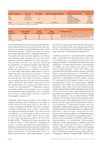
Table 7: Mortality and morbidity with GCS at admission and GOS on last follow‑up
           GCS on admission   Number       Mortality   Major residualneurodeficit  GOS on last follow‑up  Number (%)
           3‑7                11 (6.8%)       7                 1              1 (death)              13 (8.02)
           8‑12               29 (17.9%)      5                 1              2 (vegetative)           0 (0)
           13‑15             122 (75.3%)     10                 7              3 (severe disability)    0 (0)
                                                                               4 (moderate disability)  9 (5.55)
           Total                162       22 (13.58%)        9 (5.55%)         5 (good recovery)     131 (80.86)
           GCS: Glasgow Coma Scale; GOS: Glasgow Outcome Scale

           Table 8: Relationship between mortality and GCS score on admission in brain abscess
           GCS on        Total number        Total         Total        Chi‑square test
           admission       of patient      mortality     survivality
           3‑12               40              12            28          12.2 (at 5% level of significance calculated Chi‑square
           13‑15             122              10            112         test value 12.2 is greater than table value 3.84)
           Total             162              22            140
           GCS: Glasgow Coma Scale

           were isolated from abscesses of all types and at all sites,   one had one large abscess in frontal lobe and another
           whereas Enterobacteriaceae and Bacteroides spp. were   abscess in cerebritis stage on the opposite frontal lobe
           isolated from otogenic temporal lobe abscesses, which   and the rest had large paracentral lobule abscess with
           had mixed cultures. [13]  Streptococcus spp. have been   huge mass effect compressing the opposite side.
           most commonly isolated from cardiogenic abscesses. [14]
           In neonates, the most common organisms are Proteus   A lumbar puncture is contraindicated  in  patients
           and Citrobacter spp. Anaerobes are one of the most   with a suspected brain abscess because it can result
           common causative organisms in a brain abscess. [15]    in transtentorial or transforaminal herniation and
           Polymicrobial infections are common, indicating    subsequent death.  CT facilitates early detection, exact
                                                                              [26]
           the importance of using both aerobic and anaerobic   localization, accurate characterization, determination
           cultures in diagnosis. [15,16]  Cultures for acid-fast   of number, size and staging of the abscess. It also
           bacilli and fungi should be conducted in all cases   detects hydrocephalus, raised ICP, edema and
           as occasionally, intracranial tuberculosis as well as   associated infections like subdural empyema and thus
           fungal infections can present as an abscess. [17-20]  In our   helps in treatment planning. It is invaluable in the
           series, majority of the culture failed to show positive   assessment of the adequacy of treatment and sequential
           bacterial growth. More than one-third of otogenic   follow-up. An ill-defined area of low density, on plain
           and metastatic abscesses are polymicrobial (aerobic   CT, corresponds to developing necrotic center in the
                                                                            [6]
           and/or anaerobic). Bacteroides, peptostreptococcus   cerebritis stage.  In the early capsule stage, a slightly
           and fusobacterium are common anaerobes and are     hyperdense, faint ring is seen surrounding a necrotic
           sensitive to metronidazole. [21-23]  Rhinogenic abscess   hypodense center. With contrast, the ring shows thin
           is generally streptococcal. Staphylococcus is common   regular enhancement of uniform thickness and smooth
           in posttraumatic and postoperative cases. In infants   contour on its inner surface with marked perilesional
           and neonates, postmeningitic abscess is caused by   hypodense area suggestive of edema. In the late capsule
           Gram-negative organisms. [24]                      stage, the capsule is seen as a ring in plain CT. With
                                                              contrast, it shows thick enhancement gradually fading
           Clinically, brain abscess presents with features of   in delayed scans. Ring enhancement can be seen in
           rapidly expanding intracranial mass lesion that is,   the late cerebritis stage and is not an absolute evidence
           raised intracranial pressure  (ICP) in the form of   of encapsulation. [8,9]  Radiological features alone are
           constant progressive headache refractory to therapy,   inadequate to differentiate pyogenic brain abscess from
           vomiting, papilledema, focal deficits, convulsions,   fungal, nocardial or tuberculous abscess, inflammatory
           meningism and altered sensorium. The classical triad   granuloma  (tuberculoma),  neurocysticercosis,
           of headache, focal neurological deficits and fever   toxoplasmosis, metastasis, glioma, resolving haematoma,
           is found in 25% cases only.  Brain abscess occurs   infarct, hydatid cyst lymphoma and radionecrosis. [27-30]
                                     [5]
           in the younger age groups usually in the first three   However, fever, meningism, raised ESR, multilocularity,
           decades of life. [1,6]  Seizures have been reported in up   leptomeningeal or ependymal enhancement,
           to 50% of cases. [1,6,25]  The duration of symptoms is   reduction of ring enhancement in delayed scan and
           usually < 2 weeks, with rapid onset and progression.   finding of gas within the lesion favor a diagnosis of
           Immunocompromised patients may have an insidious   abscess.  Positive labeling in radionuclide imaging
                                                                      [9]
           onset.  Three patients in this series had bladder and   with III-Indium labeled leukocytes, C-reactive protein,
                [6]
           bowel incontinence; one had tubercular abscess in   99m TC-hexamethylpropylene amine oxime leukocyte
           third ventricular floor with hydrocephalus, second   scintigraphy, diffusion weighted MRI, Thallium-201


            158                                              Neuroimmunol Neuroinflammation | Volume 2 | Issue 3 | July 15, 2015  Neuroimmunol Neuroinflammation | Volume 2 | Issue 3 | July 15, 2015                              159