
Page 171 - Read Online
P. 171
chemotherapy or target agents, [15,16,20] it is irreplaceable the cannulas. Dexamethasone sodium phosphate was
in the treatment of NM, despite of some reported diluted from 1 mL (5 mg) to 5 mL with physiological
adverse reactions. [21,22] We acquire some clinical saline and then slowly injected into the subarachnoid
experience about how to minimize the side effect and space. During the injection, dexamethasone sodium
how to institute the course of treatment. phosphate was mixed with drawing back CSF repeatedly.
Methotrexate was diluted to 5 mL and then injected the
METHODS same way dexamethasone was treated.
Inclusion criteria Data collection
Subjects were required to present with the clinical signs The patients’ characteristics and treatment information at
and symptoms consistent with NM, including headache, the diagnosis of NM were obtained in the medical record
confusion, cranial and spinal nerve involvement, nausea from the Second Hospital of Hebei Medical University.
and vomit. CSF (200 μL) was collected from a lumbar Survival data, subsequent therapeutic schedule, and side
puncture and it was centrifuged in (650 rpm) for 4 min effects following discharge were obtained by making
using Slide Centrifuge (Shandon Cytospin 4, Thermo). the phone calls to ask whether there is paralysis, severe
Cell slides were May-Grunwald-Giemsa stained for vomiting, headache within 48 h after intrathecal injection,
5 min then phosphate buffers was added and incubated or the symptoms of bone marrow suppression, such as
for 10 min, followed by gentle rinsing with running fever, infection, and low blood cell count. Overall survival
water. The stained cell slides were observed under the was calculated from the diagnosis of NM.
microscope (Oil immersion lens ×1000, Olympus DP72).
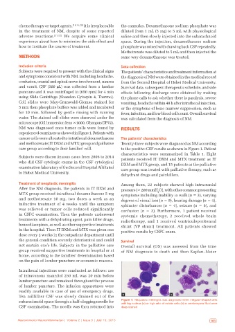
NM was diagnosed once tumor cells were found by RESULTS
experienced examiners as showed in Figure 1. Patients with
cancer cells were allocated to intrathecal dexamethasone The patients’ characteristics
and methotrexate (IT DXM and MTX) group and palliative Twenty-three subjects were diagnosed as NM according
care group according to their families’ will. to the positive CSF results as shown in Figure 1. Patient
characteristics were summarized in Table 1. Eight
Subjects were discontinuous cases from 2006 to 2014 patients received IT DXM and MTX treatment as IT
who did CSF cytologic exams in the CSF cytological DXM and MTX group, and 15 patients as the palliative
examination laboratory of the Second Hospital Affiliated care group was treated with palliative therapy, such as
to Hebei Medical University.
dehydrant drugs and painkillers.
Treatment of neoplastic meningitis Among them, 22 subjects showed high intracranial
After the NM diagnosis, the patients in IT DXM and pressure (> 200 mmH O), with other common presenting
2
MTX group received intrathecal dexamethasone 5 mg symptoms including inability to walk (n = 3), varying
and methotrexate 10 mg, two doses a week as an degrees of visual loss (n = 9), hearing damage (n = 4),
inductive treatment of 4 weeks until the symptom sphincter disturbances (n = 4), seizure (n = 6), and
was relieved or tumor cells reduced significantly confusion (n = 3). Furthermore, 1 patient received
in CSFC examination. Then the patients underwent systemic chemotherapy, 2 received whole brain
treatments with a dehydrating agent, pain killer drugs, radiotherapy, and 1 received ventriculoperitoneal
benzodiazepines, as well as other supportive treatments shunt (VP shunt) treatment. All patients showed
in the hospital. Then IT DXM and MTX was given one positive results by CSFC exam.
dose every 2 weeks in the outpatient department until
the general condition severely deteriorated and could Survival
not sustain one’s life. Subjects in the palliative care Overall survival (OS) was assessed from the time
group received supportive treatments in hospital or at of NM diagnosis to death and then Kaplan–Meier
home, according to the families’ determination based
on the pain of lumber puncture or economic reasons.
Intrathecal injections were conducted as follows: use
of intravenous mannitol 250 mL was 20 min before
lumbar puncture and remained throughout the process
of lumber puncture. The infusion apparatuses were
readily available in case of use of emergency drugs.
Ten milliliter CSF was slowly drained out of the a b
subarachnoid space through a half-clogging needle for Figure 1: Neoplastic meningitis was diagnosed when irregular‑shaped cells
with big nucleus (a) or high ratio of mitotic cells (b) in cerebrospinal fluid were
CSF examination. The needle was then returned into deep‑stained
162 Neuroimmunol Neuroinflammation | Volume 2 | Issue 3 | July 15, 2015 Neuroimmunol Neuroinflammation | Volume 2 | Issue 3 | July 15, 2015 163