
Page 176 - Read Online
P. 176
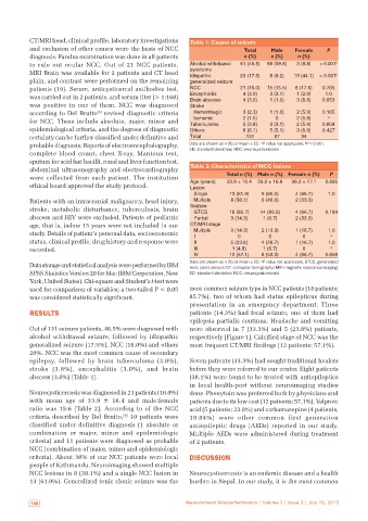
CT/MRI head, clinical profile, laboratory investigations   Table 1: Causes of seizure
           and exclusion of other causes were the basis of NCC                  Total    Male   Female     P
           diagnosis. Fundus examination was done in all patients               n (%)    n (%)   n (%)
           to rule out ocular NCC. Out of 21 NCC patients,    Alcohal withdrawal   61 (46.5)  58 (59.8)  3 (8.8)  < 0.001 b
           MRI Brain was available for 2 patients and CT head   syndrome       23 (17.5)  8 (8.2)  15 (44.1)  < 0.001 b
                                                              Idiopathic
           plain, and contrast were performed on the remaining   generalized seizure
           patients (19). Serum, anticysticercal antibodies test,   NCC        21 (16.0)  15 (15.5)  6 (17.6)  0.765
                                                                                                          1.0
           was carried out in 2 patients, and serum titer (> 1:160)   Encephalitis  4 (3.0)  3 (3.1)  1 (2.9)  0.053
                                                                                4 (3.0)
                                                                                        1 (1.0)
                                                              Brain abscess
                                                                                                 3 (8.8)
           was positive in one of them. NCC was diagnosed     Stroke
           according to Del Brutto  revised diagnostic criteria   Hemorrhagic   3 (2.3)  1 (1.0)  2 (5.9)  0.165
                                [9]
                                                                Ischemic
                                                                                          0
                                                                                                           a
           for NCC. These include absolute, major, minor and   Tuberculoma      2 (1.5)  3 (3.1)  2 (5.9)  0.604
                                                                                5 (3.8)
                                                                                                 2 (5.9)
           epidemiological criteria, and the degrees of diagnostic   Others     8 (6.1)  5 (5.1)  3 (8.8)  0.427
           certainty can be further classified under definitive and   Total      131      97      34
                                                                                                    b
                                                                                      a
           probable diagnosis. Reports of electroencephalography,   Data are shown as n (%) or mean ± SD.  P value not applicable;  P < 0.001.
           complete blood count, chest X-ray, Mantoux test,   SD: standard deviation; NCC: neurocysticercosis
           sputum for acid fast bacilli, renal and liver function test,
           abdominal ultrasonography and electrocardiography   Table 2: Characteristics of NCC lesions
           were  collected  from  each  patient.  The  institution        Total n (%) Male n (%)  Female n (%)  P
           ethical board approved the study protocol.          Age (years)  33.9 ± 16.4  33.0 ± 16.6  36.3 ± 17.1  0.685
                                                               Lesion
                                                                Single     13 (61.9)  9 (60.0)   4 (66.7)  1.0
           Patients with an intracranial malignancy, head injury,   Multiple  8 (38.1)  6 (40.0)  2 (33.3)
           stroke, metabolic disturbance, tuberculosis, brain   Seizure    18 (85.7)  14 (93.3)  4 (66.7)  0.184
                                                                GTCS
           abscess and HIV were excluded. Patients of pediatric   Partial   3 (14.3)  1 (6.7)    2 (33.3)
           age, that is, below 15 years were not included in our   CT/MRI stage
           study. Details of patient’s personal data, socioeconomic   Multiple  3 (14.3)  2 (13.3)  1 (16.7)  1.0
                                                                                                   0
                                                                                                            a
                                                                I
                                                                              0
                                                                                        0
           status, clinical profile, drug history and response were   II    5 (23.8)  4 (26.7)   1 (16.7)  1.0
           recorded.                                            III         1 (4.8)   1 (6.7)      0        a
                                                                IV         12 (57.1)  8 (53.3)   4 (66.7)  0.659
                                                                                      a
           Data storage and statistical analysis were performed by IBM   Data are shown as n (%) or mean ± SD.  P value not applicable. GTCS: generalized
                                                              tonic clonic seizure; CT: computer tomography; MRI: magnetic resonance imaging;
           SPSS Statistics Version 20 for Mac (IBM Corporation, New   SD: standard deviation; NCC: neurocysticercosis
           York, United States). Chi-square and Student’s t-test were
           used for comparison of variables; a two-tailed P < 0.05   most common seizure type in NCC patients (18 patients;
           was considered statistically significant.          85.7%), two of whom had status epilepticus during
                                                              presentation in an emergency department. Three
           RESULTS                                            patients (14.3%) had focal seizure, one of them had
                                                              epilepsia partialis continua. Headache and vomiting
           Out of 131 seizure patients, 46.5% were diagnosed with   were observed in 7 (33.3%) and 5 (23.8%) patients,
           alcohol withdrawal seizure, followed by idiopathic   respectively [Figure 1]. Calcified stage of NCC was the
           generalized seizure (17.5%), NCC (16.0%) and others   most frequent CT/MRI findings (12 patients; 57.1%).
           20%. NCC was the most common cause of secondary
           epilepsy, followed by brain tuberculoma  (3.8%),   Seven patients (33.3%) had sought traditional healers
           stroke  (3.8%),  encephalitis  (3.0%),  and  brain   before they were referred to our center. Eight patients
           abscess (3.0%) [Table 1].                          (38.1%) were found to be treated with antiepileptics
                                                              in  local  health-post  without  neuroimaging  studies
           Neurocysticercosis was diagnosed in 21 patients (16.0%)   done. Phenytoin was preferred both by physicians and
           with mean age of 33.9  ±  16.4 and male:female     patients due to its low cost (12 patients; 57.1%). Valproic
           ratio was 15:6 [Table  2]. According to of the NCC   acid (5 patients; 23.8%) and carbamazepine (4 patients,
           criteria described by Del Brutto,  10 patients were   19.04%) were other common first generation
                                         [9]
           classified under definitive diagnosis (1 absolute or   antiepileptic drugs  (AEDs) reported in our study.
           combination or major, minor and epidemiologic      Multiple AEDs were administered during treatment
           criteria) and 11 patients were diagnosed as probable   of 2 patients.
           NCC (combination of major, minor and epidemiologic
           criteria). About 30% of our NCC patients were local   DISCUSSION
           people of Kathmandu. Neuroimaging showed multiple
           NCC lesions in 8 (38.1%) and a single NCC lesion in   Neurocysticercosis is an endemic disease and a health
           13 (61.9%). Generalized tonic clonic seizure was the   burden in Nepal. In our study, it is the most common


            168                                              Neuroimmunol Neuroinflammation | Volume 2 | Issue 3 | July 15, 2015  Neuroimmunol Neuroinflammation | Volume 2 | Issue 3 | July 15, 2015                              169