
Page 180 - Read Online
P. 180
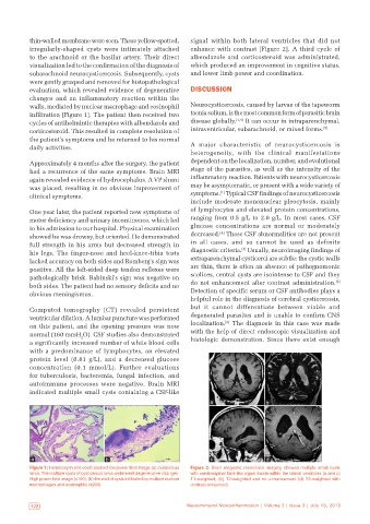
thin-walled membrane were seen. These yellow-spotted,   signal within both lateral ventricles that did not
           irregularly-shaped cysts were intimately attached   enhance  with  contrast [Figure  2].  A  third  cycle  of
           to the arachnoid or the basilar artery. Their direct   albendazole and corticosteroid was administrated,
           visualization led to the confirmation of the diagnosis of   which produced an improvement in cognitive status,
           subarachnoid neurocysticercosis. Subsequently, cysts   and lower limb power and coordination.
           were gently grasped and removed for histopathological
           evaluation, which revealed evidence of degenerative   DISCUSSION
           changes and an inflammatory reaction within the
           walls, mediated by nuclear macrophage and eosinophil   Neurocysticercosis, caused by larvae of the tapeworm
           infiltration [Figure 1]. The patient then received two   taenia solium, is the most common form of parasitic brain
           cycles of antihelmintic therapies with albendazole and   disease globally. [1,4]  It can occur in intraparenchymal,
           corticosteroid. This resulted in complete resolution of   intraventricular, subarachnoid, or mixed forms. [5]
           the patient’s symptoms and he returned to his normal
           daily activities.                                  A major characteristic of neurocysticercosis is
                                                              heterogeneity, with the clinical manifestations
           Approximately 4 months after the surgery, the patient   dependent on the localization, number, and evolutional
           had a recurrence of the same symptoms. Brain MRI   stage of the parasites, as well as the intensity of the
           again revealed evidence of hydrocephalus. A VP shunt   inflammatory reaction. Patients with neurocysticercosis
           was placed, resulting in no obvious improvement of   may be asymptomatic, or present with a wide variety of
                                                                        [1]
           clinical symptoms.                                 symptoms.  Typical CSF findings of neurocysticercosis
                                                              include moderate mononuclear pleocytosis, mainly
           One year later, the patient reported new symptoms of   of lymphocytes and elevated protein concentrations,
           motor deficiency and urinary incontinence, which led   ranging from 0.5 g/L to 2.0 g/L. In most cases, CSF
           to his admission to our hospital. Physical examination   glucose concentrations are normal or moderately
                                                                        [4]
           showed he was drowsy, but oriented. He demonstrated   decreased.  These CSF abnormalities are not present
           full strength in his arms but decreased strength in   in all cases, and so cannot be used as definite
                                                                               [3]
           his legs.  The  finger-nose and  heel-knee-tibia tests   diagnostic criteria.  Usually, neuroimaging findings of
           lacked accuracy on both sides and Romberg’s sign was   extraparenchymal cysticerci are subtle: the cystic walls
           positive. All the left-sided deep tendon reflexes were   are thin, there is often an absence of pathognomonic
           pathologically brisk. Babinski’s sign was negative on   scolices, central cysts are isointense to CSF and they
                                                                                                             [3]
           both sides. The patient had no sensory deficits and no   do not enhancement after contrast administration.
           obvious meningismus.                               Detection of specific serum or CSF antibodies plays a
                                                              helpful role in the diagnosis of cerebral cysticercosis,
                                                              but  it cannot  differentiate  between  viable  and
           Computed tomography  (CT) revealed persistent
           ventricular dilation. A lumbar puncture was performed   degenerated parasites and is unable to confirm CNS
                                                                          [6]
           on this patient, and the opening pressure was now   localization.  The diagnosis in this case was made
           normal (160 mmH O). CSF studies also demonstrated   with the help of direct endoscopic visualization and
                           2
           a significantly increased number of white blood cells   histologic demonstration. Since there exist enough
           with  a  predominance  of  lymphocytes,  an  elevated
           protein level  (0.81  g/L), and a decreased glucose
           concentration  (0.1 mmol/L). Further evaluations
           for tuberculosis, bacteremia, fungal infection, and
           autoimmune  processes  were negative. Brain  MRI
           indicated multiple small cysts containing a CSF-like
                                                                     a                 b


           a                       b                                 c                 d
           Figure 1: Hematoxylin and eosin stained low power field image (a) cysticercus   Figure 2: Brain magnetic resonance imaging showed multiple small cysts
           larva. The multiple cysts of cysticercus larva underwent degenerative changes.   with cerebrospinal fluid‑like signal inside within the lateral ventricles (a and c)
           High power field image (x100); (b) the wall of cysts infiltrated by multiple nuclear   T1‑weighted,  (b) T2‑weighted  and no  enhancement  (d) T2‑weighted  with
           macrophages and eosinophils (x200)                 contrast‑enhanced)


            172                                              Neuroimmunol Neuroinflammation | Volume 2 | Issue 3 | July 15, 2015  Neuroimmunol Neuroinflammation | Volume 2 | Issue 3 | July 15, 2015                              173