
Page 183 - Read Online
P. 183
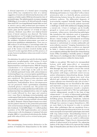
A clinical impression of a frontal space occupying   case  include  the  butterfly  configuration,  forniceal
           lesion  (SOL) was considered in view of a history   thickening and features of a “mass-effect” with a clinical
           suggestive of mesial and orbitofrontal dysfunction with   presentation akin to a butterfly glioma; prominent
           suspicion of white-matter (WM) involvement in view of   differentiating features being the enhancement and
           pyramidal signs. The initial magnetic resonance imaging   perfusion patterns. The differential diagnosis of
           (MRI) [Figure 1] revealed an ill-defined butterfly shaped   acute-subacute acquired “butterfly lesions” involving
           intra axial SOL involving bilateral frontal lobes as well   the corpus callosum as seen in the patient represents
           as anterior insula with predominant involvement of   a challenge in itself with multimodal imaging playing
           subcortical deep WM and extension into bilateral caudate   a crucial role. A  host of etiologies can be broadly
           nuclei, genu and the anterior 1/3 of body of corpus   grouped as: tumors such as glioma, lymphoma and
           callosum. Moderate mass-effect over bilateral frontal   metastasis; inflammatory demyelinating pathologies
           horns of lateral ventricles was observed. The lesion   like tumefactive MS; infections such as progressive
           was hyperintense on T2-weighted and hypointense on   multifocal leukoencephalopathy and Whipple’s
           T1-weighted images with mild diffusion restriction over   disease; toxins leading to disseminated necrotizing
           the periphery. Peripheral enhancement was noted on   leukoencephalopathy, e.g.  intrathecal or systemic
           intravenous gadolinium administration. There was no   exposure to methotrexate or cytosine arabinoside and
           MR evidence of calcification or hemorrhage within the   acute radiation necrosis.  Imaging characteristics that
                                                                                    [1]
           lesion. MR spectroscopy (MRS) from the intermediate   potentially differentiate these conditions are depicted
           part of the lesion revealed elevated choline peak,   in Table  1. Clinico-radiological presentation of a
           reduced N-acetyl aspartate (NAA) and the presence of   non-neoplastic pathology like tumefactive demyelinating
           lactate; however perfusion values were noted to be low   lesion (TDL) resembling a butterfly glioma is fraught
           in the lesion.                                     with chance of a misdiagnosis (due to heterogeneity of
                                                              imaging characteristics within the lesion itself).
           On admission, the patient was noted to develop rapidly
           progressive encephalopathy with features of raised   Unlike in our patient, TDL tend to be circumscribed
           intracranial pressure in the form of bradycardia,   lesions with mild mass-effect or vasogenic
           hypersomnolence and hypertension. With the imaging   edema.  These typically involve the supra-tentorial
                                                                     [2]
           consideration of an intermediate-high grade  SOL   WM although they may extend to involve the cortical
           and gliomatosis cerebri high on the cards in view   gray matter with gyral edema. In a large series of
           of a butterfly-patterned lesion with calloso-forniceal   168 patients with biopsy confirmed central nervous
           thickening [Figure 1a], a neuronavigation-guided biopsy   system inflammatory demyelinating disease, frontal
           and as an alternative a frontal decompressive procedure   and parietal subcortical regions were most often
           was considered. The frozen-section specimen and    affected and a butterfly configuration involving the
           histopathology report [Figure 2] was consistent with                                              [3]
           tumefactive demyelination hence decompression      corpus callosum was observed in only 12% of cases.
           was not performed. Such a presentation mimicking   Forniceal thickening was not described in this series
           a butterfly glioma is extremely rare. The patient was   and represents a unique observation in our patient
           treated with pulse methyl predisolone followed by oral   as this deviated the impression towards a neoplastic
           prednisolone that was administered in a dose of 1 mg/kg   etiology. Approximately half of TDL have pathological
           for 8  weeks followed by slow taper and cessation   contrast enhancement, usually in the form of ring
           over 1 year. Prior to discharge evoked potentials, and   enhancement. [2,3]  A variety of intracranial pathologies
           cerebrospinal fluid studies including oligoclonal bands   can present as a ring-enhancing lesion (REL) on MRI,
           were negative thereby making multiple sclerosis (MS)   including  glioma,  metastasis,  lymphoma,  radiation
           less likely. Dramatic clinical improvement was noted,   necrosis, infarct, abscess and tumefactive demyelination.
           and the patient returned to the premorbid personality   Although less common in typical demyelination, REL
           with normal neuropsychological performance 3 months   are more likely to be biopsied in order to exclude
           into treatment. Serial MRI [Figure 3] verified gradual   these  other  pathologies  that  are  mandatory  from  a
           resolution of the WM hyperintensities and contrast   treatment and prognostication point of view. In a recent
           enhancement with development of minimal bifrontal   series among the most prevalent pathologies associated
           atrophy. MRS at 1 year showed reduction in the choline   with ring enhancement, demyelinating lesions of
           peak with reduced NAA and no evident lactate peak.  MS constituted a small number (6%) and patterns of
                                                              T2-weighted hypointensity are useful to differentiate
           DISCUSSION                                         between pathologies.  Figure 1b and c demonstrate this
                                                                                 [4]
                                                              pattern of enhancement in the patient. The enhancing
           The neuroimaging characteristics distinctive in the   portion of the ring is believed to represent the leading
           pattern of tumefactive demyelination in the index   edge of demyelination and thus favors the WM side of


   174  Neuroimmunol Neuroinflammation | Volume 2 | Issue 3 | July 15, 2015  Neuroimmunol Neuroinflammation | Volume 2 | Issue 3 | July 15, 2015   175