
Page 933 - Read Online
P. 933
Shaikh et al. Mini-invasive Surg 2020;4:89 I http://dx.doi.org/10.20517/2574-1225.2020.97 Page 5 of 19
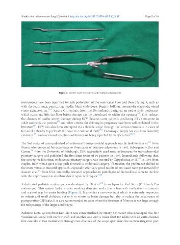
Figure 4. MINOP shaft instrument with multiple attachments
instruments have been described for safe perforation of the ventricular floor and then dilating it, such as
with the leucotome, puncturing needle, blunt endoscope, Fogarty balloon, monopolar electrode, wired
[22]
stone extractor, etc. . Andre Grotenhuis from the Netherlands designed an endoscopic perforator
[23]
which sucks and lifts the floor before forceps can be introduced to widen the opening . This reduces
the chances of basilar artery damage during ETV. Success score systems predicting ETV’s outcome in
[24]
adult and pediatric patients and other criteria for defining its prognosis have been well explained in the
[25]
literature . ETV has also been attempted via a flexible scope through the lamina terminalis in cases of
[26]
technical difficulty to perforate the floor via traditional route . Endoscopic biopsy has also been favorably
[27]
evaluated , and occasional resections of tumors are being reported by many centers [28,29] .
[30]
The first series of cases published of endonasal transsphenoidal approach was by Jankowski et al. from
France who presented his experience in three cases of pituitary adenomas in 1992. Subsequently, Jho and
[31]
Carrau from the University of Pittsburgh, USA successfully used nasal endoscopes for transsphenoidal
pituitary surgery and published the first large series of 50 patients in 1997. Immediately following that,
[32]
the concept of functional endoscopic pituitary surgery was mooted by Cappabianca et al. in 1998 from
Naples, Italy, which gave a big push forward to endonasal surgery. Thereafter, the preference shifted to
the more versatile binostril approach, especially after very good results of 800 cases were put forward by
Kassam et al. from USA. Gradually, extended approaches to pathologies of the skullbase came to the fore
[33]
with the improvement in skullbase defect repair techniques [34,35] .
[36]
A dedicated pediatric endoscope was developed by Oi et al. from Japan for Karl Storz (Oi Handy Pro
endoscope). This system had a smaller working diameter and a 2-mm lens with malleable instruments
and a pistol grip for easier holding [Figure 5]. It provides a narrower tract which is extremely important
in infants and small children, not only to minimize brain damage but also to reduce the occurrence of
postoperative CSF leaks. It is also recommended in cases where the foramen of Monroe is not large enough
for safe passage of the larger adult scope.
Pediatric Lotta system from Karl Storz was conceptualized by Henry Schroeder who developed this HD
visualization scope with narrow shaft and another one with a wider shaft for adults with an extra channel
that can take in two instruments through two channels of the scope apart from the suction-irrigation port