
Page 932 - Read Online
P. 932
Page 4 of 19 Shaikh et al. Mini-invasive Surg 2020;4:89 I http://dx.doi.org/10.20517/2574-1225.2020.97
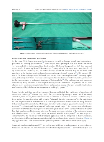
Figure 3. Decq scope and its tip with multiple channels and malleable instruments which can pass through it
Endoscopes and endoscopic procedures
In the 1990s, Claris Corporation was the first to come out with endoscope guided ventricular catheter
[12]
placement for treating hydrocephalus . These scopes were lightweight, thin with outer diameter of
1.14 mm, and able to be introduced into shunt catheters. Medtronic Company from USA then came out
with a similar functioning NeuroPEN endoscope. Correspondingly, slit tip catheters were introduced
by Medtronic and Codman (USA) for ventriculoscopic placement. However, they did not attain wide
[13]
acceptance as the literature consists of experiences mentioning only small case series . This was probably
[14]
due to the absence of any discernible benefit over routine shunt catheter placements , relatively higher
costs, and suboptimal vision. However, neurosurgeons have not been deterred from probing avenues for
[15]
further improvements in endoscopic treatment of hydrocephalus . The multipurpose ventriculoscope
described by Henry Schroeder in 2008 helps in tackling not only obstructive CSF pathways but the extra
channel allows also intraventricular lesion biopsy and resection, among other uses ably aided by the then
newly developed high definition (HD) visualization and display system [16,17] .
Bauer, Hellwig, and their team from Marburg, Germany published their eight years of experience of
[18]
stereotactic endoscopy wherein they used it for cystic cerebral pathologies, intracerebral hematoma
[19]
evacuation, brain abscess, third ventriculostomy, and retrieval of ventricular catheters. Axel Perneczky
from Mainz, Germany is credited with bringing “minimally invasive neurosurgery” to the mainstream
in 1998 by greater use of narrower (MINOP, Aesculap) endoscopes in ventricles and using them for
indications beyond hydrocephalus. He brought stereotaxy and navigation guidance in endoscopy to the
[20]
forefront and developed the concept of “endoscope guided surgery” for cases such as colloid cysts.
Endoscope assisted microneurosurgery was the next stage in the mid-1990s and innovations to attain the
best dual imaging were highly sought after. Axel Perneczky proposed projection of the endoscopic images
[1]
into a head mounted LCD device which was not routinely available in that period . His most important
contribution was the concept of “keyhole surgical approaches” with the integration of these visualization
methods to the skullbase and development of specially designed shaft instruments for dissection [Figure 4],
clip applicators, and a table mounted endoscope holding device to aid bimanual endoscopic surgery.
Endoscopic third ventriculostomy (ETV) is one of the most widely performed procedures in neuroendoscopy
[21]
today and its results have been validated worldwide for hydrocephalus . Several techniques and