
Page 930 - Read Online
P. 930
Page 2 of 19 Shaikh et al. Mini-invasive Surg 2020;4:89 I http://dx.doi.org/10.20517/2574-1225.2020.97
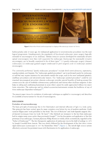
Figure 1. Maximilian Nitze’s urethroscope kept on display at the endoscopy museum at Vienna
hydrocephalus only 25 years ago. Its widespread application to intraventricular procedures was the next
logical progression. Simultaneously, the popularity of functional endoscopic sinus surgery logically
extended to neurosurgery of the skullbase. Tubular retractor systems developed for minimally invasive
spinal neurosurgery were then well supported by endoscopy. Endoscopy for minimally invasive
[1]
neurosurgery can be broadly considered to be of three types : (1) purely endoscopic surgery (channel
endoscopy); (2) endoscope guided surgery (endonasal or port surgery); and (3) endoscope assisted
microsurgery.
The commonly performed “purely endoscopic procedures” include third ventriculostomy, septostomy,
aqueductoplasty, and biopsies. The “endoscopic guided procedures” can be performed purely by endoscopy
as well but may require assistance by instruments outside the scope, such as for most endonasal pituitary,
skullbase procedures and intraventricular tumors. The “endoscopy assisted procedure” could be any
standard microsurgical procedure wherein endoscopy provides special benefits of looking around the
corners as in the case of vestibular schwannomas, epidermoid, and various other skullbase surgeries. It also
helps in aiding and confirming hemostasis in areas which cannot be easily approached without too much
brain retraction. The endoscope and its related accessories/instruments remain the backbone of any of
[2,3]
these endoscope-dependent techniques .
The present paper traces the evolution of endoscopic techniques as applied to neurosurgery and describes
the available armamentarium for the aid of neurosurgeons.
DISCUSSION
Evolution of neuroendoscopy
The basic principle of endoscopy lies in the illumination and internal reflection of light in a body cavity.
This principle has been worked upon by many scientists even before the era of modern medicine. Greek
scientist Hippocrates’ work published in the book “The Art of Medicine” and Arab-Spanish surgeon Abu-
al-Qasim’s techniques from the book “Al-Tasrif” (The Method) are testament to the fact that endoscopy
[4]
had its origins many years earlier than previously thought . For his description and application of the first
prototype of an endoscope, German physician Philip Bozzini is widely, albeit contentiously, regarded as the
[5]
“Father of Endoscopy” . The first therapeutic application of endoscopy was in the field of urology in 1873
by Joseph Grunfeld from Austria. This was closely followed by the development of the first direct-vision
[6]
rigid endoscopes (cystoscope) in 1877 by Maximilian Nitze [Figure 1]. The inbuilt light source system
effectively corrected the persistent issues with illumination in the application of endoscopy.