
Page 931 - Read Online
P. 931
Shaikh et al. Mini-invasive Surg 2020;4:89 I http://dx.doi.org/10.20517/2574-1225.2020.97 Page 3 of 19
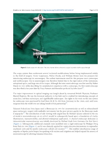
Figure 2. Gaab scope with obturator. The side channel allows ultrasonic surgical aspirator shaft to pass through
The scope system then underwent several technical modifications before being implemented widely
in the field of surgery. Victor Lespinasse, Walter Dandy, and William Mixter were the pioneers for
introducing endoscopy in neurosurgery. The earliest instruments used for this purpose were cystoscopes
and urethroscopes. Use in neurosurgery was therefore limited due to the rigid nature of the instrument,
suboptimal optics, and large size of the scopes. Although the term ventriculoscopy was first used by Walter
Dandy in 1922 while describing his unsatisfactory experience with a cystoscope, the first ventriculoscope
[7]
was described a few years later by Tracy Putnam and thereafter perfected by John Scarrf .
The major improvement in optical imaging was bought about by renowned British Physicist, Professor
Harold Hopkins. He was the foremost authority in his field and is credited for introducing concepts of
zoom lens, rod-lens endoscopes, and rigid/flexible endoscopes. The rights to his work on the lens system
for endoscope were purchased by Karl Storz SE & Co. KG from Germany in the 1960s, and, until now,
[8]
surgeons from the world over are taking benefit of this partnership .
Takanori Fukushima from Japan used a fiberscope in 1973 for intraventricular as well as subarachnoid
space endoscopic surgery with malleable instruments but the poor picture quality in the fiberscope made
it unpopular . The introduction of side viewing wide angled lens by Michael Apuzzo ushered in the era
[9]
of modern neuroendoscopy, an era which would be subsequently based upon a foundation of clarity,
illumination, maneuverability, and allowed widespread application. A channel endoscope dedicated to
intraventricular neuroendoscopy was initially developed by Michael Gaab from Germany (for Karl Storz)
[Figure 2]. Subsequently, additional channels were modified onto a rigid endoscope by Philippe Decq
from Paris in 1996 [Figure 3], and it was clinically applied for ventriculocystocisternostomy in suprasellar
arachnoid cysts and for purely endoscopic colloid cyst excision [10,11] . This enabled simultaneous usage of
unipolar or bipolar probe biopsy forcepsalong with suction and irrigation and helped expand the armory of
neuroendoscopy by allowing bimanual dissection.