
Page 937 - Read Online
P. 937
Shaikh et al. Mini-invasive Surg 2020;4:89 I http://dx.doi.org/10.20517/2574-1225.2020.97 Page 9 of 19
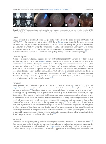
Figure 9. A: MRI T2W coronal section of giant suprasellar arachnoid cyst; B: coagulation of cyst wall by utilizing thulium LASER
endoscopically; C: cyst wall after application of the LASER; D: perforated cyst wall; E: intracystic visualization after ventriculocystostomy
LASER
LASER application in neuroendoscopy has gradually evolved from the initial use of Nd:YAG and KTP
LASERr [45,46] to the more recent introduction of Thulium [Figure 9], considered to be more precise and
efficacious than its predecessors. Hypothalamic hamartoma disconnection by using thulium represents a
[47]
good example of LASER replacing the conventional coagulation technique in neurosurgery . To combat
the issues of damage to healthy brain tissue, LASER now consists of pretreated, carbon coated, diode fiber
[48]
tip to prevent deeper neurovascular structures from getting damaged with the dissipating energy .
Ultrasonic aspirator
[49]
Results of endoscopic ultrasonic aspirator use were first published in 2008 by Oertel et al. . Since then, it
has been used for intraventricular [Figure 10] and paraventricular lesions along with thulium LASER for
[50]
hemostasis . For endonasal surgeries, special thin and long tip ultrasonic aspirators can be used (Both
ultastrasonic aspirators by Soering, Germany). We have found ultrasonic aspirators to beuseful but severe
limitation can be faced due to repeated blockages and because it can only be used presently with Gaab
endoscope channel. Barrow Institute from Arizona, USA introduced a variable aspirator and described
[44]
its use for endoscopic resection of hypothalamic hamartoma in 2006 . Numerous case series have since
described the utility of a multipurpose side cutting aspirator (NICO, Myriad, USA) in neuroendoscopy
especially in patients with intraventricular tumors [51-54] .
Navigation
Image guidance in neuroendoscopy has become a vital tool for planning and trajectory guidance
[55]
[Figure 11] and has been proven to add value to some if not all procedures . A global survey of 235
[56]
neurosurgeons in 2012 found that image guidance was used always in conjunction with intraventricular
endoscopy by approximately 17% of participants, especially for tumor biopsy, resection, and cyst
fenestration. When it came to endoscopic skullbase surgery, image guidance was used for all cases by 24%
of respondents, and more so for recurrent and complex skullbase anatomy cases. Navigation has also been
effectively combined with virtual endoscopy, i.e., magnetic resonance ventriculoscopy, to help reduce
[57]
chances of damage to critical structures during endoscopic surgery . Technically, the tool has advanced
over years by overcoming the initial shortcoming of head fixation considered imperative for many years
to achieve accuracy. There has since been development of navigation system with face mask (Stryker) and
electromagnetic system by companies such as Medtronic, Brainlab, etc. Neither system requires the head to
be fixed with pins during surgery. Today, neuronavigation also plays a significant role in simulation training
for endoscopy in cadavers as well as synthetic models [58,59] .
Ultrasound
Ultrasound for navigation guiding neuroendoscopy procedures was described as early as the 1990s [60,61] .
This can be very useful if a child has an open fontanelle and is undergoing endoscopic treatment for
complex hydrocephalus or multiloculated cysts [Figure 12] and is effectively used by many surgeons as
[62]
an alternative to MR guided procedures . Intracranial application of ultrasound probes concurrent with