
Page 938 - Read Online
P. 938
Page 10 of 19 Shaikh et al. Mini-invasive Surg 2020;4:89 I http://dx.doi.org/10.20517/2574-1225.2020.97
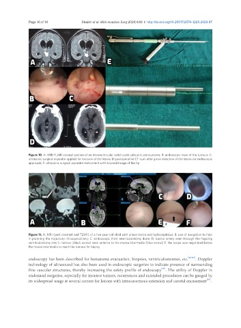
Figure 10. A: MRI FLAIR coronal section of an intraventricular solid cystic pilocytic astrocytoma; B: endoscopic view of the tumour; C:
ultrasonic surgical aspirator applied for excision of the lesion; D: postoperative CT scan after gross resection of the lesion via endoscopic
approach; E: ultrasonic surgical aspirator instrument with zoomed image of the tip
Figure 11. A: MRI (post contrast and T2WI) of a five-year-old child with pineal lesion and hydrocephalus; B: use of navigation to help
in planning the trajectory intraoperatively; C: endoscopic third ventriculostomy done; D: basilar artery seen through the flapping
ventriculostomy site; E: tumour (black arrow) seen anterior to the massa intermedia (blue arrow); F: the scope was negotiated below
the massa intermedia to reach the tumour for biopsy
endoscopy has been described for hematoma evacuation, biopsies, ventriculostomies, etc. [63-65] . Doppler
technology of ultrasound has also been used in endoscopic surgeries to indicate presence of surrounding
[66]
fine vascular structures, thereby increasing the safety profile of endoscopy . The utility of Doppler in
endonasal surgeries, especially for invasive tumors, recurrences and extended procedures can be gauged by
[67]
its widespread usage at several centers for lesions with intracavernous extension and carotid encasement .