
Page 939 - Read Online
P. 939
Shaikh et al. Mini-invasive Surg 2020;4:89 I http://dx.doi.org/10.20517/2574-1225.2020.97 Page 11 of 19
Figure 12. A: MRI T2W axial section showing multiloculated hydrocephalus; B: ultrasound image for guidance of septostomy; C:
navigation image showing successful septostomy with passing of catheter to the opposite ventricle
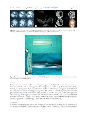
Figure 13. A: the tubular retractor designed by Yadav et al. [70] in 2011. Longitudinal cut allows the retractor to be folded onto itself; B: the
small size prevents a large cortical opening
Retractors
Tubular retractors represent another avenue of augmenting the minimally invasive nature of neuroendoscopy.
For intraventricular lesions, a transparent cylindrical port was developed by Daniel Prevedello, Amin
[68]
Kassam, and their group . Many series have been published elaborating on transparent sheath retractor
[69]
use for ventricular tumors including syringe ports . We have used simple transparent tubes for some
[70]
years now for deep seated lesion excision, while Yadav et al. used it with a small slit to reduce pressure
on surrounding brain [Figure 13]. Tubular retractors have also been modified for use as a nasal retractor
in skullbase endoscopic surgery . Even though the field of vision is proven to be better with microscope
[71]
[72]
assisted surgery than neuroendoscopy , many surgeons still favor these retractors.
Hemostats
Hemostasis aiding endoscopic surgery still relies greatly on conventional and long existing methods such
as copious warm irrigation, absorbable gelatin sponges (Gelfoam from Baxter), and oxidized regenerated