
Page 22 - Read Online
P. 22
Page 16 of 25 Singh et al. Mini-invasive Surg. 2025;9:28 https://dx.doi.org/10.20517/2574-1225.2024.75
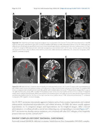
Figure 22. ENB, Kadish Group B (nasal cavity and paranasal sinus involvement). (A) Coronal CT shows soft-tissue attenuation in the left
superior nasal cavity and ethmoid sinus; (B) Coronal T2-weighted MRI shows a mildly hyperintense tumor in the left nasal cavity and
ethmoid sinus. Peripheral entrapped fluid (red arrow) shows marked hyperintensity compared with the tumor (yellow arrow); (C) Post-
contrast T1-weighted MRI demonstrates relatively homogeneous solid enhancement of the tumor (yellow arrow), whereas the
entrapped fluid shows only peripheral enhancement (red arrow). ENB: Esthesioneuroblastoma; CT: computed tomography; MRI:
magnetic resonance imaging.
Figure 23. ENB, Kadish Group C (extension beyond nasal cavity and paranasal sinuses). (A) Coronal CT shows soft-tissue attenuation in
the bilateral nasal cavities and ethmoid sinuses, with dehiscence of the cribriform plate (red arrow); (B) Coronal T2-weighted MRI
shows a sinonasal tumor extending intracranially through the right cribriform plate (red arrow). T2-hyperintense trapped fluid is seen in
the right maxillary sinus, along with a rounded T2-hypointense lesion at the sinus floor (yellow arrow); (C) Post-contrast T1-weighted
image shows relatively homogeneous solid enhancement of the sinonasal tumor, with early intracranial dural invasion (red arrow) and
an enhancing satellite lesion at the floor of the right maxillary sinus (yellow arrow). ENB: Esthesioneuroblastoma; CT: computed
tomography; MRI: magnetic resonance imaging.
On CT, NUT carcinoma demonstrates aggressive features such as bony erosion, hyperostosis, avid contrast
enhancement, intralesional mineralization, and orbital invasion. On MRI, the tumor usually appears
hypointense on T1-weighted images and hyperintense on T2-weighted images. After contrast
[41]
administration, it is enhanced with a central non-enhancing area, corresponding to necrosis [Figure 25] .
NUT carcinoma may develop osseous metastases, and compared with other SCC subtypes, it less frequently
presents with non-necrotic cervical LAD .
[41]
SWI/SNF COMPLEX-DEFICIENT SINONASAL CARCINOMAS
Previously termed SMARCB1-deficient carcinoma, Switch/Sucrose Non-Fermentable (SWI/SNF) complex-