
Page 27 - Read Online
P. 27
Singh et al. Mini-invasive Surg. 2025;9:28 https://dx.doi.org/10.20517/2574-1225.2024.75 Page 21 of 25
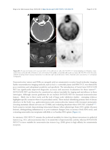
Figure 29. 49-year-old female with recurrent AdCC of the ethmoid air cells. (A) FDG PET/CT demonstrating an infiltrative, trans-
spatial mass centered in the ethmoid air cells with marked FDG uptake, compatible with local recurrence; (B) FDG PET/CT in the same
patient revealing distant osseous metastasis in a sclerotic right sacral ala lesion. AdCC: Adenoid cystic carcinoma; CT: computed
tomography.
Neuroendocrine tumors and ENBs are uniquely avid on somatostatin receptor-based molecular imaging.
Earlier neuroendocrine imaging methods, such as Ind111-octreotide planar and SPECT/CT, were limited by
poor resolution and suboptimal sensitivity and specificity. The introduction of Ga68/Cu64-DOTATATE
PET has significantly improved diagnostic accuracy and anatomic localization for these tumors .
[61]
DOTATATE PET can therefore be considered an adjunct modality for staging and surveillance in these
histologies. Although current guidelines do not include DOTATE PET for sinonasal neuroendocrine
tumors - likely due to their rarity and the lack of large-scale validation studies - emerging evidence
highlights specific scenarios where it is particularly useful. These include identifying primary sites of disease
elsewhere in the body (e.g., gastroenteropancreatic neuroendocrine tumors with sinonasal metastasis),
detecting metastatic disease not seen on CT/MRI, and evaluating situations where FDG PET is limited [62,63] .
Such scenarios include characterizing intracranial disease (where physiologic brain FDG uptake obscures
lesions), distinguishing inflammatory or post-treatment changes from recurrence (both FDG-avid), and
evaluating neuroendocrine neoplasms with little or no baseline FDG uptake [Figure 30].
In summary, FDG PET/CT remains the preferred modality for detecting distant metastases in epithelial
tumors (e.g., SCC, adenocarcinoma) due to its sensitivity to hypermetabolic activity, whereas DOTATATE
PET/CT is more suitable for neuroendocrine tumors (e.g., ENB) given its high affinity for somatostatin
receptors.