
Page 25 - Read Online
P. 25
Singh et al. Mini-invasive Surg. 2025;9:28 https://dx.doi.org/10.20517/2574-1225.2024.75 Page 19 of 25
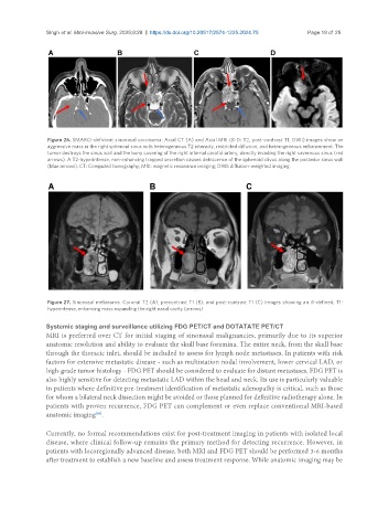
Figure 26. SMARCI-deficient sinonasal carcinoma: Axial CT (A) and Axial MRI (B-D: T2, post-contrast T1, DWI) images show an
aggressive mass in the right sphenoid sinus with heterogeneous T2 intensity, restricted diffusion, and heterogeneous enhancement. The
tumor destroys the sinus wall and the bony covering of the right internal carotid artery, directly invading the right cavernous sinus (red
arrows). A T2-hyperintense, non-enhancing trapped secretion causes dehiscence of the sphenoid clivus along the posterior sinus wall
(blue arrows). CT: Computed tomography; MRI: magnetic resonance imaging; DWI: diffusion-weighted imaging.
Figure 27. Sinonasal melanoma. Coronal T2 (A), precontrast T1 (B), and post-contrast T1 (C) images showing an ill-defined, T1-
hyperintense, enhancing mass expanding the right nasal cavity (arrows).
Systemic staging and surveillance utilizing FDG PET/CT and DOTATATE PET/CT
MRI is preferred over CT for initial staging of sinonasal malignancies, primarily due to its superior
anatomic resolution and ability to evaluate the skull base foramina. The entire neck, from the skull base
through the thoracic inlet, should be included to assess for lymph node metastases. In patients with risk
factors for extensive metastatic disease - such as multistation nodal involvement, lower cervical LAD, or
high-grade tumor histology - FDG PET should be considered to evaluate for distant metastases. FDG PET is
also highly sensitive for detecting metastatic LAD within the head and neck. Its use is particularly valuable
in patients where definitive pre-treatment identification of metastatic adenopathy is critical, such as those
for whom a bilateral neck dissection might be avoided or those planned for definitive radiotherapy alone. In
patients with proven recurrence, FDG PET can complement or even replace conventional MRI-based
[49]
anatomic imaging .
Currently, no formal recommendations exist for post-treatment imaging in patients with isolated local
disease, where clinical follow-up remains the primary method for detecting recurrence. However, in
patients with locoregionally advanced disease, both MRI and FDG PET should be performed 3-6 months
after treatment to establish a new baseline and assess treatment response. While anatomic imaging may be