
Page 21 - Read Online
P. 21
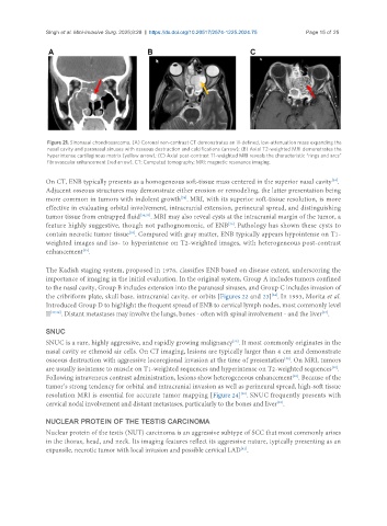
Singh et al. Mini-invasive Surg. 2025;9:28  https://dx.doi.org/10.20517/2574-1225.2024.75  Page 15 of 25


                Figure 21. Sinonasal chondrosarcoma. (A) Coronal non-contrast CT demonstrates an ill-defined, low-attenuation mass expanding the
                nasal cavity and paranasal sinuses with osseous destruction and calcifications (arrow); (B) Axial T2-weighted MRI demonstrates the
                hyperintense cartilaginous matrix (yellow arrow); (C) Axial post-contrast T1-weighted MRI reveals the characteristic “rings and arcs”
                fibrovascular enhancement (red arrow). CT: Computed tomography; MRI: magnetic resonance imaging.

               On CT, ENB typically presents as a homogeneous soft-tissue mass centered in the superior nasal cavity .
                                                                                                       [34]
               Adjacent osseous structures may demonstrate either erosion or remodeling, the latter presentation being
               more common in tumors with indolent growth . MRI, with its superior soft-tissue resolution, is more
                                                         [34]
               effective in evaluating orbital involvement, intracranial extension, perineural spread, and distinguishing
               tumor tissue from entrapped fluid [34,35] . MRI may also reveal cysts at the intracranial margin of the tumor, a
               feature highly suggestive, though not pathognomonic, of ENB . Pathology has shown these cysts to
                                                                       [36]
               contain necrotic tumor tissue . Compared with gray matter, ENB typically appears hypointense on T1-
                                         [35]
               weighted images and iso- to hyperintense on T2-weighted images, with heterogeneous post-contrast
               enhancement .
                           [35]
               The Kadish staging system, proposed in 1976, classifies ENB based on disease extent, underscoring the
               importance of imaging in the initial evaluation. In the original system, Group A includes tumors confined
               to the nasal cavity, Group B includes extension into the paranasal sinuses, and Group C includes invasion of
                                                                                     [34]
               the cribriform plate, skull base, intracranial cavity, or orbits [Figures 22 and 23] . In 1993, Morita et al.
               Introduced Group D to highlight the frequent spread of ENB to cervical lymph nodes, most commonly level
               II [37,38] . Distant metastases may involve the lungs, bones - often with spinal involvement - and the liver .
                                                                                                    [37]
               SNUC
                                                                        [39]
               SNUC is a rare, highly aggressive, and rapidly growing malignancy . It most commonly originates in the
               nasal cavity or ethmoid air cells. On CT imaging, lesions are typically larger than 4 cm and demonstrate
                                                                                        [40]
               osseous destruction with aggressive locoregional invasion at the time of presentation . On MRI, tumors
               are usually isointense to muscle on T1-weighted sequences and hyperintense on T2-weighted sequences .
                                                                                                       [40]
               Following intravenous contrast administration, lesions show heterogeneous enhancement . Because of the
                                                                                           [40]
               tumor’s strong tendency for orbital and intracranial invasion as well as perineural spread, high-soft tissue
               resolution MRI is essential for accurate tumor mapping [Figure 24] . SNUC frequently presents with
                                                                           [40]
                                                                                      [39]
               cervical nodal involvement and distant metastases, particularly to the bones and liver .
               NUCLEAR PROTEIN OF THE TESTIS CARCINOMA
               Nuclear protein of the testis (NUT) carcinoma is an aggressive subtype of SCC that most commonly arises
               in the thorax, head, and neck. Its imaging features reflect its aggressive nature, typically presenting as an
                                                                           [41]
               expansile, necrotic tumor with local invasion and possible cervical LAD .