
Page 17 - Read Online
P. 17
Singh et al. Mini-invasive Surg. 2025;9:28 https://dx.doi.org/10.20517/2574-1225.2024.75 Page 11 of 25
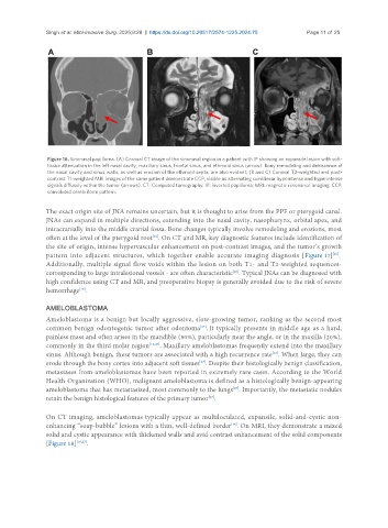
Figure 16. Sinonasal papilloma. (A) Coronal CT image of the sinonasal region in a patient with IP showing an expansile lesion with soft-
tissue attenuation in the left nasal cavity, maxillary sinus, frontal sinus, and ethmoid sinus (arrow). Bony remodeling and dehiscence of
the nasal cavity and sinus walls, as well as erosion of the ethmoid septa, are also evident; (B and C) Coronal T2-weighted and post-
contrast T1-weighted MRI images of the same patient demonstrate CCP, visible as alternating curvilinear hypointense and hyperintense
signals diffusely within the tumor (arrows). CT: Computed tomography; IP: inverted papilloma; MRI: magnetic resonance imaging; CCP:
convoluted cerebriform pattern.
The exact origin site of JNA remains uncertain, but it is thought to arise from the PPF or pterygoid canal.
JNAs can expand in multiple directions, extending into the nasal cavity, nasopharynx, orbital apex, and
intracranially into the middle cranial fossa. Bone changes typically involve remodeling and erosions, most
often at the level of the pterygoid root . On CT and MR, key diagnostic features include identification of
[16]
the site of origin, intense hypervascular enhancement on post-contrast images, and the tumor’s growth
pattern into adjacent structures, which together enable accurate imaging diagnosis [Figure 17] .
[26]
Additionally, multiple signal flow voids within the lesion on both T1- and T2-weighted sequences-
corresponding to large intralesional vessels - are often characteristic . Typical JNAs can be diagnosed with
[26]
high confidence using CT and MR, and preoperative biopsy is generally avoided due to the risk of severe
hemorrhage .
[16]
AMELOBLASTOMA
Ameloblastoma is a benign but locally aggressive, slow-growing tumor, ranking as the second most
[27]
common benign odontogenic tumor after odontoma . It typically presents in middle age as a hard,
painless mass and often arises in the mandible (80%), particularly near the angle, or in the maxilla (20%),
commonly in the third molar region [16,27] . Maxillary ameloblastomas frequently extend into the maxillary
[16]
sinus. Although benign, these tumors are associated with a high recurrence rate . When large, they can
erode through the bony cortex into adjacent soft tissues . Despite their histologically benign classification,
[27]
metastases from ameloblastomas have been reported in extremely rare cases. According to the World
Health Organization (WHO), malignant ameloblastoma is defined as a histologically benign-appearing
[27]
ameloblastoma that has metastasized, most commonly to the lungs . Importantly, the metastatic nodules
[27]
retain the benign histological features of the primary tumor .
On CT imaging, ameloblastomas typically appear as multiloculated, expansile, solid-and-cystic non-
enhancing “soap-bubble” lesions with a thin, well-defined border . On MRI, they demonstrate a mixed
[16]
solid and cystic appearance with thickened walls and avid contrast enhancement of the solid components
[Figure 18] [16,27] .