
Page 14 - Read Online
P. 14
Page 8 of 25 Singh et al. Mini-invasive Surg. 2025;9:28 https://dx.doi.org/10.20517/2574-1225.2024.75
Figure 11. MRI tissue characterization. MRI: Magnetic resonance imaging.
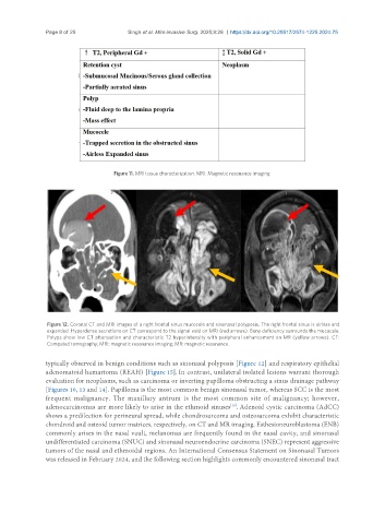
Figure 12. Coronal CT and MRI images of a right frontal sinus mucocele and sinonasal polyposis. The right frontal sinus is airless and
expanded. Hyperdense secretions on CT correspond to the signal void on MRI (red arrows). Bony deficiency surrounds the mucocele.
Polyps show low CT attenuation and characteristic T2 hyperintensity with peripheral enhancement on MR (yellow arrows). CT:
Computed tomography; MRI: magnetic resonance imaging; MR: magnetic resonance.
typically observed in benign conditions such as sinonasal polyposis [Figure 12] and respiratory epithelial
adenomatoid hamartoma (REAH) [Figure 15]. In contrast, unilateral isolated lesions warrant thorough
evaluation for neoplasms, such as carcinoma or inverting papilloma obstructing a sinus drainage pathway
[Figures 10, 13 and 14]. Papilloma is the most common benign sinonasal tumor, whereas SCC is the most
frequent malignancy. The maxillary antrum is the most common site of malignancy; however,
adenocarcinomas are more likely to arise in the ethmoid sinuses . Adenoid cystic carcinoma (AdCC)
[19]
shows a predilection for perineural spread, while chondrosarcoma and osteosarcoma exhibit characteristic
chondroid and osteoid tumor matrices, respectively, on CT and MR imaging. Esthesioneuroblastoma (ENB)
commonly arises in the nasal vault, melanomas are frequently found in the nasal cavity, and sinonasal
undifferentiated carcinoma (SNUC) and sinonasal neuroendocrine carcinoma (SNEC) represent aggressive
tumors of the nasal and ethmoidal regions. An International Consensus Statement on Sinonasal Tumors
was released in February 2024, and the following section highlights commonly encountered sinonasal tract