
Page 18 - Read Online
P. 18
Page 12 of 25 Singh et al. Mini-invasive Surg. 2025;9:28 https://dx.doi.org/10.20517/2574-1225.2024.75
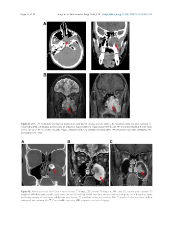
Figure 17. JNA. (A) Axial (left) and coronal (right) non-contrast CT images, and (B) coronal T2-weighted (left) and post-contrast T1-
weighted (right) MRI images, demonstrate an expansile, hyperenhancing mass arising from the left PPF and extending into the left nasal
cavity (arrows). JNA: Juvenile nasopharyngeal angiofibroma; CT: computed tomography; MRI: magnetic resonance imaging; PPF:
pterygopalatine fossa.
Figure 18. Ameloblastoma. (A) Coronal non-contrast CT image, (B) Coronal T2-weighted MRI, and (C) coronal post-contrast T1-
weighted MRI show an expansile cystic mass (arrow, B) involving the left maxillary alveolus and extending into the left maxillary sinus.
Avid enhancement of the internal solid component (arrow, C) is evident on the post-contrast MRI. The lesion is also associated with an
unerupted tooth (arrow, A). CT: Computed tomography; MRI: magnetic resonance imaging.