
Page 20 - Read Online
P. 20
Page 14 of 25 Singh et al. Mini-invasive Surg. 2025;9:28 https://dx.doi.org/10.20517/2574-1225.2024.75
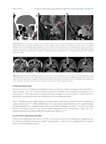
Figure 19. SNSCC. (A) Coronal CT image of the sinonasal cavity shows a destructive soft-tissue mass centered in the left maxillary
antrum with erosion of the sinus wall, nasal cavity, ethmoid complex, orbit, and anterior skull base (arrow); (B) Coronal T2-weighted
MRI demonstrates intermediate signal intensity of the tumor and loss of the hypointense zone, indicating dural invasion (arrow); (C)
Coronal post-contrast T1-weighted MRI shows nodular tumor extension through the anterior skull base defect with hyperenhancing
thickening of the overlying dura (arrow). SNSCC: Sinonasal squamous cell carcinoma; CT: computed tomography; MRI: magnetic
resonance imaging.
Figure 20. AdCC. From left to right: axial precontrast T1, axial T2, and three axial post-contrast T1 images. AdCC demonstrates invasion
of the right skull base with sclerosis of the pterygoid and sphenoid bones (white), and perineural invasion involving the PPF, Vidian canal
(blue), foramen ovale (yellow), foramen rotundum (green), inferior orbital fissure (orange), superior orbital fissure (red), orbital apex
(red), and ventral cavernous sinus (red). AdCC: Adenoid cystic carcinoma; PPF: pterygopalatine fossa.
CHONDROSARCOMA
[16]
Chondrosarcoma is a malignant cartilaginous tumor commonly arising in the long bones and pelvis .
Approximately 10% occur in the head and neck, with the maxillary sinus being the most frequent site of
involvement [16,33] . The peak incidence is observed between the ages of 40 and 60 years [16,33] . These tumors are
typically slow-growing and often cause deformation of surrounding structures.
On CT, chondrosarcomas usually appear as soft tissue masses with matrix calcifications and heterogeneous
contrast enhancement [16,33] . MRI typically shows low to intermediate signal intensity on T1-weighted images
and high intensity on T2-weighted images in the non-calcified cartilaginous regions . Calcified areas often
[33]
produce blooming artifacts. Post-contrast MRI demonstrates the characteristic “rings and arcs”
enhancement pattern associated with fibrovascular tissue [Figure 21] [16,33] .
OLFACTORY NEUROBLASTOMA
Olfactory neuroblastoma, also known as ENB, is a rare neural crest-derived malignancy originating from
[34]
the olfactory epithelium of the nasal cavity . Imaging plays a critical role in mapping both locoregional
tumor extent and metastatic disease.