
Page 23 - Read Online
P. 23
Singh et al. Mini-invasive Surg. 2025;9:28 https://dx.doi.org/10.20517/2574-1225.2024.75 Page 17 of 25
Figure 24. SNUC. (A) Coronal CT image of the paranasal sinus shows diffuse soft-tissue opacification of the right nasal cavity and
ethmoid complex, with erosion of the ethmoid septa. The maxillary sinus is also opacified with lower attenuation; (B) Coronal T2-
weighted MRI of the same patient shows intermediate tumor signal (red arrow). Maxillary sinus opacification appears hyperintense,
consistent with retained secretions (yellow arrow); (C) Post-contrast T1-weighted MRI shows heterogeneous tumor enhancement (red
arrow). The maxillary sinus demonstrates enhancing thickened mucosa, consistent with inflammatory disease (yellow arrow). SNUC:
Sinonasal undifferentiated carcinoma; CT: computed tomography; MRI: magnetic resonance imaging.
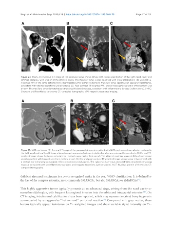
Figure 25. NUT carcinoma. (A) Coronal CT image of the paranasal sinuses in a patient with NUT carcinoma shows a lesion centered in
the right nasal cavity with soft-tissue attenuation and aggressive features, including both bone erosion and hyperostosis; (B) Coronal T2-
weighted image shows the tumor as isointense relative to gray matter (red arrow). The adjacent maxillary sinus exhibits a hyperintense
signal consistent with trapped secretions (yellow arrow); (C) Coronal post-contrast T1-weighted image shows tumor enhancement with
a central non-enhancing component reflecting necrosis (red arrow). The right maxillary sinus demonstrates peripheral enhancing
mucosa, consistent with an inflammatory process and trapped secretions (yellow arrow). NUT: Nuclear protein of the testis; CT:
computed tomography.
deficient sinonasal carcinoma is a newly recognized entity in the 2022 WHO classification. It is defined by
the loss of the complex subunits, most commonly SMARCB1, but also SMARCA2 or SMARCA4 .
[25]
This highly aggressive tumor typically presents at an advanced stage, arising from the nasal cavity or
nasoethmoidal region, with frequent locoregional invasion into the orbits and intracranial extension . On
[42]
CT imaging, intralesional calcifications have been reported, which may represent retained bony fragments
accompanied by an aggressive “hair-on-end” periosteal reaction . Compared with gray matter, these
[42]
lesions typically appear isointense on T1-weighted images and show variable signal intensity on T2-