
Page 26 - Read Online
P. 26
Page 20 of 25 Singh et al. Mini-invasive Surg. 2025;9:28 https://dx.doi.org/10.20517/2574-1225.2024.75
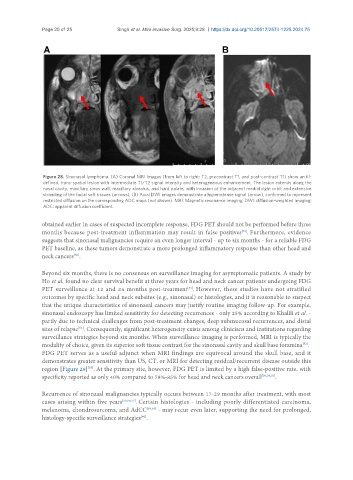
Figure 28. Sinonasal lymphoma. (A) Coronal MRI images (from left to right: T2, precontrast T1, and post-contrast T1) show an ill-
defined, trans-spatial lesion with intermediate T1/T2 signal intensity and heterogeneous enhancement. The lesion extends along the
nasal cavity, maxillary sinus wall, maxillary alveolus, and hard palate, with invasion of the adjacent medial right orbit and extensive
stranding of the facial soft tissues (arrows); (B) Axial DWI images demonstrate a hyperintense signal (arrow), confirmed to represent
restricted diffusion on the corresponding ADC maps (not shown). MRI: Magnetic resonance imaging; DWI: diffusion-weighted imaging;
ADC: apparent diffusion coefficient.
obtained earlier in cases of suspected incomplete response, FDG PET should not be performed before three
months because post-treatment inflammation may result in false positives . Furthermore, evidence
[49]
suggests that sinonasal malignancies require an even longer interval - up to six months - for a reliable FDG
PET baseline, as these tumors demonstrate a more prolonged inflammatory response than other head and
neck cancers .
[50]
Beyond six months, there is no consensus on surveillance imaging for asymptomatic patients. A study by
Ho et al. found no clear survival benefit at three years for head and neck cancer patients undergoing FDG
PET surveillance at 12 and 24 months post-treatment . However, these studies have not stratified
[51]
outcomes by specific head and neck subsites (e.g., sinonasal) or histologies, and it is reasonable to suspect
that the unique characteristics of sinonasal cancers may justify routine imaging follow-up. For example,
sinonasal endoscopy has limited sensitivity for detecting recurrences - only 25% according to Khalili et al. -
partly due to technical challenges from post-treatment changes, deep submucosal recurrences, and distal
sites of relapse . Consequently, significant heterogeneity exists among clinicians and institutions regarding
[52]
surveillance strategies beyond six months. When surveillance imaging is performed, MRI is typically the
modality of choice, given its superior soft tissue contrast for the sinonasal cavity and skull base foramina .
[52]
FDG PET serves as a useful adjunct when MRI findings are equivocal around the skull base, and it
demonstrates greater sensitivity than US, CT, or MRI for detecting residual/recurrent disease outside this
[53]
region [Figure 29] . At the primary site, however, FDG PET is limited by a high false-positive rate, with
specificity reported as only 40% compared to 78%-85% for head and neck cancers overall [50,54,55] .
Recurrence of sinonasal malignancies typically occurs between 17-29 months after treatment, with most
cases arising within five years [50,56,57] . Certain histologies - including poorly differentiated carcinoma,
melanoma, chondrosarcoma, and AdCC [58,59] - may recur even later, supporting the need for prolonged,
histology-specific surveillance strategies .
[60]