
Page 16 - Read Online
P. 16
Page 10 of 25 Singh et al. Mini-invasive Surg. 2025;9:28 https://dx.doi.org/10.20517/2574-1225.2024.75
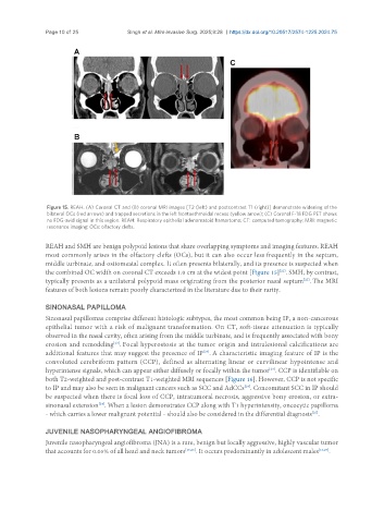
Figure 15. REAH. (A) Coronal CT and (B) coronal MRI images [T2 (left) and postcontrast T1 (right)] demonstrate widening of the
bilateral OCs (red arrows) and trapped secretions in the left frontoethmoidal recess (yellow arrow); (C) Coronal F-18 FDG PET shows
no FDG-avid signal in this region. REAH: Respiratory epithelial adenomatoid hamartoma; CT: computed tomography; MRI: magnetic
resonance imaging; OCs: olfactory clefts.
REAH and SMH are benign polypoid lesions that share overlapping symptoms and imaging features. REAH
most commonly arises in the olfactory clefts (OCs), but it can also occur less frequently in the septum,
middle turbinate, and ostiomeatal complex. It often presents bilaterally, and its presence is suspected when
the combined OC width on coronal CT exceeds 1.0 cm at the widest point [Figure 15] . SMH, by contrast,
[21]
[22]
typically presents as a unilateral polypoid mass originating from the posterior nasal septum . The MRI
features of both lesions remain poorly characterized in the literature due to their rarity.
SINONASAL PAPILLOMA
Sinonasal papillomas comprise different histologic subtypes, the most common being IP, a non-cancerous
epithelial tumor with a risk of malignant transformation. On CT, soft-tissue attenuation is typically
observed in the nasal cavity, often arising from the middle turbinate, and is frequently associated with bony
erosion and remodeling . Focal hyperostosis at the tumor origin and intralesional calcifications are
[23]
[23]
additional features that may suggest the presence of IP . A characteristic imaging feature of IP is the
convoluted cerebriform pattern (CCP), defined as alternating linear or curvilinear hypointense and
hyperintense signals, which can appear either diffusely or focally within the tumor . CCP is identifiable on
[24]
both T2-weighted and post-contrast T1-weighted MRI sequences [Figure 16]. However, CCP is not specific
[24]
to IP and may also be seen in malignant cancers such as SCC and AdCCs . Concomitant SCC in IP should
be suspected when there is focal loss of CCP, intratumoral necrosis, aggressive bony erosion, or extra-
sinonasal extension . When a lesion demonstrates CCP along with T1 hyperintensity, oncocytic papilloma
[24]
- which carries a lower malignant potential - should also be considered in the differential diagnosis .
[25]
JUVENILE NASOPHARYNGEAL ANGIOFIBROMA
Juvenile nasopharyngeal angiofibroma (JNA) is a rare, benign but locally aggressive, highly vascular tumor
that accounts for 0.05% of all head and neck tumors [16,26] . It occurs predominantly in adolescent males [16,27] .