
Page 12 - Read Online
P. 12
Page 6 of 25 Singh et al. Mini-invasive Surg. 2025;9:28 https://dx.doi.org/10.20517/2574-1225.2024.75
Figure 7. (A) Coronal CT showing the characteristic “ring and arc” appearance of a chondroid matrix in chondrosarcoma; (B) CT
demonstrating “sunburst” periostitis and osteoid matrix typical of osteosarcoma. CT: Computed tomography.
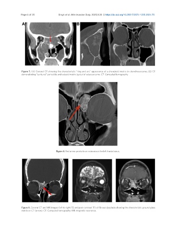
Figure 8. Red arrow points to an osteoma in the left frontal sinus.
Figure 9. Coronal CT and MR images (left to right: T2 and post-contrast T1) of fibrous dysplasia showing the characteristic ground-glass
matrix on CT (arrow). CT: Computed tomography; MR: magnetic resonance.