
Page 10 - Read Online
P. 10
Page 4 of 25 Singh et al. Mini-invasive Surg. 2025;9:28 https://dx.doi.org/10.20517/2574-1225.2024.75
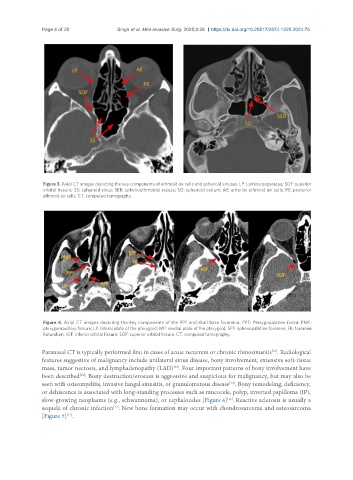
Figure 3. Axial CT images depicting the key components of ethmoid air cells and sphenoid sinuses. LP: Lamina papyracea; SOF: superior
orbital fissure; SS: sphenoid sinus; SER: sphenoethmoidal recess; SO: sphenoid ostium; AE: anterior ethmoid air cells; PE: posterior
ethmoid air cells; CT: computed tomography.
Figure 4. Axial CT images depicting the key components of the PPF and skull base foramina. PPF: Pterygopalatine fossa; PMF:
pterygomaxillary fissure; LP: lateral plate of the pterygoid; MP: medial plate of the pterygoid; SPF: sphenopalatine foramen; FR: foramen
Rotundum; IOF: inferior orbital fissure; SOF: superior orbital fissure; CT: computed tomography.
Paranasal CT is typically performed first in cases of acute recurrent or chronic rhinosinusitis . Radiological
[12]
features suggestive of malignancy include unilateral sinus disease, bony involvement, extensive soft-tissue
mass, tumor necrosis, and lymphadenopathy (LAD) . Four important patterns of bony involvement have
[12]
[12]
been described : Bony destruction/erosion is aggressive and suspicious for malignancy, but may also be
[12]
seen with osteomyelitis, invasive fungal sinusitis, or granulomatous disease . Bony remodeling, deficiency,
or dehiscence is associated with long-standing processes such as mucocele, polyp, inverted papilloma (IP),
[12]
slow-growing neoplasms (e.g., schwannoma), or cephaloceles [Figure 6] . Reactive sclerosis is usually a
sequela of chronic infection . New bone formation may occur with chondrosarcoma and osteosarcoma
[12]
[Figure 7] .
[12]