
Page 96 - Read Online
P. 96
Selvakumar et al. IMT post LDLT-report of first case
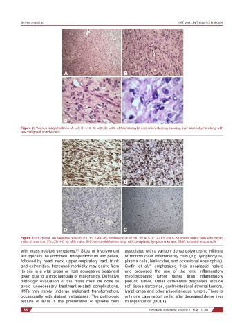
Figure 2: Various magnifications (A: ×4; B: ×10; C: ×20; D: ×40) of haematoxylin and eosin staining showing liver parenchyma along with
few malignant spindle cells
Figure 3: IHC panel. (A) Negative result of IHC for SMA; (B) positive result of IHC for ALK-1l; (C) IHC for C-Kit shows tumor cells with mitotic
index of less than 5%; (D) IHC for MIB index. IHC: immunohistochemistry; ALK: anaplastic lymphoma kinase; SMA: smooth muscle actin
with mass related symptoms. Sites of involvement associated with a variably dense polymorphic infiltrate
[5]
are typically the abdomen, retroperitoneum and pelvis, of mononuclear inflammatory cells (e.g. lymphocytes,
followed by head, neck, upper respiratory tract, trunk plasma cells, histiocytes, and occasional eosinophils).
and extremities. Increased morbidity may derive from Coffin et al. emphasized their neoplastic nature
[6]
its site in a vital organ or from aggressive treatment and proposed the use of the term inflammatory
given due to a misdiagnosis of malignancy. Definitive myofibroblastic tumor rather than inflammatory
histologic evaluation of the mass must be done to pseudo tumor. Other differential diagnoses include
avoid unnecessary treatment-related complications. soft tissue sarcomas, gastrointestinal stromal tumors,
IMTs may rarely undergo malignant transformation, lymphomas and other miscellaneous tumors. There is
occasionally with distant metastases. The pathologic only one case report so far after deceased donor liver
feature of IMTs is the proliferation of spindle cells transplantation (DDLT).
88 Hepatoma Research ¦ Volume 3 ¦ May 17, 2017