
Page 95 - Read Online
P. 95
Selvakumar et al. IMT post LDLT-report of first case
confirmed IMT with anaplastic lymphoma kinase activity. was also seen near the mass. Two other small tumors
She received ceritinibas chemotherapy for 1 year. At of approximately 2 cm each were seen attached to
1.5 years of follow-up, the child is recurrence free. serosa of small bowel. All the macroscopically visible
tumors were completely removed. HPE revealed IMT
CASE REPORT of the peritoneum with very mild malignant potential.
Immunohistochemistry (IHC) was positive for
Our patient was a 1-year-old female child from anaplastic lymphoma kinase (ALK) antibodies [Figures
Pakistan. She had decompensated chronic liver 2 and 3]. Postoperative period was uneventful. She
disease secondary to EHBA. She underwent primary was discharged in a hemodynamically stable condition
LDLT at the age of 6 months at our center. Her mother on postoperative day 10. After tumor board discussion,
was the donor. She had uneventful post transplantation ceritinib was given as adjuvant therapy in view of ALK
recovery. At 3 months post liver transplantation (LT), positivity. At 18 months follow-up, patient is recurrence
routine USG revealed solid masses in the pelvis and free with good graft function.
in the sub hepatic region. Trucut biopsy of both the
lesions revealed mesenchymal neoplasm. There DISCUSSION
was no evidence of malignancy. There was gradual
increase in size of tumor on follow-up scans over the The risk of de novo malignancy following LT is
next 3 months [Figure 1A and B]. She reported back to significantly higher than that of the general population.
our center for further evaluation of the intra-abdominal Skin, hematological, and colon cancers are common
tumors. Review of previous biopsy slides and fresh de novo malignancies after LT. Immunosuppression
trucut biopsies done at our center were inconclusive. A plays a major role in oncogenesis in the transplant
tumor board decision was made for surgical excision to population. Other risk factors are hepatitis C virus
[1]
get further information about the tumor and also relieve infection, smoking, alcoholic cirrhosis, and sun
the mass effect in the abdomen. Elective laparotomy exposure. [2]
was planned. In laparotomy, a 15-cm lesion was seen
in right sub diaphragmatic region pushing the hepatic IMTs are one of the rare groups of post-transplant
flexure and graft liver which was also adherent to malignancies. More than 200 cases have been
diaphragm [Figure 1C and D]. Another 7-cm firm mass described so far. Majority of them are after HSCT. [3,4]
was found adherent to omentum, proximal jejunum IMTs occur earlier in the post-transplant period ranging
and jejunal mesentery. The tumor was removed along between 3 months and 2.5 years. Presenting complaints
with a segment of jejunum. Minimal loculated ascites of patients are usually fever, weight loss and pain, along
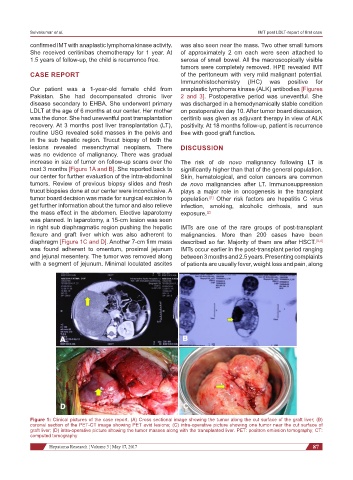
Figure 1: Clinical pictures of the case report. (A) Cross sectional image showing the tumor along the cut surface of the graft liver; (B)
coronal section of the PET-CT image showing PET avid lesions; (C) intra-operative picture showing one tumor near the cut surface of
graft liver; (D) intra-operative picture showing the tumor masses along with the transplanted liver. PET: positron emission tomography; CT:
computed tomography
Hepatoma Research ¦ Volume 3 ¦ May 17, 2017 87