
Page 307 - Read Online
P. 307
Chok et al. Pheochromocytoma mimicking primary liver cancer
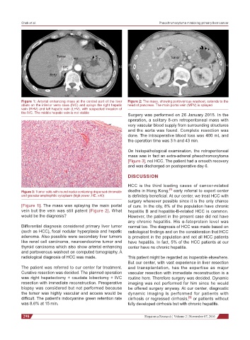
Figure 1: Arterial enhancing mass at the central part of the liver Figure 2: The mass, showing portovenous washout, extends to the
abuts on the inferior vena cava (IVC) and splays the right hepatic head of pancreas. The main portal vein (MPV) is splayed
vein (RHV) and left hepatic vein (LHV), with suspected invasion of
the IVC. The middle hepatic vein is not visible
Surgery was performed on 26 January 2015. In the
operation, a solitary 8-cm retroperitoneal mass with
very vascular blood supply from surrounding structures
and the aorta was found. Complete resection was
done. The intraoperative blood loss was 400 mL and
the operation time was 3 h and 43 min.
On histopathological examination, the retroperitoneal
mass was in fact an extra-adrenal pheochromocytoma
[Figure 3], not HCC. The patient had a smooth recovery
and was discharged on postoperative day 6.
DISCUSSION
HCC is the third leading cause of cancer-related
[1]
Figure 3: Tumor cells with round nuclei containing dispersed chromatin deaths in Hong Kong; early referral to expert center
and granular amphophilic cytoplasm (high power, HE, ×40) is definitely beneficial. At our center, we treat HCC with
surgery whenever possible since it is the only chance
[Figure 1]. The mass was splaying the main portal of cure. In the city, 8% of the population have chronic
vein but the vein was still patent [Figure 2]. What hepatitis B and hepatitis-B-related HCC is common.
would be the diagnosis? However, the patient in the present case did not have
any chronic hepatitis. His α-fetoprotein level was
Differential diagnosis considered primary liver tumor normal too. The diagnosis of HCC was made based on
(such as HCC), focal nodular hyperplasia and hepatic radiological findings and on the consideration that HCC
adenoma. Also possible were secondary liver tumors is prevalent in the population and not all HCC patients
like renal cell carcinoma, neuroendocrine tumor and have hepatitis. In fact, 5% of the HCC patients at our
thyroid carcinoma which also show arterial enhancing center have no chronic hepatitis.
and portovenous washout on computed tomography. A
radiological diagnosis of HCC was made. This patient might be regarded as inoperable elsewhere.
But our center, with vast experience in liver resection
The patient was referred to our center for treatment. and transplantation, has the expertise as major
Curative resection was decided. The planned operation vascular resection with immediate reconstruction is a
was right hepatectomy + caudate lobectomy + IVC routine here. Therefore surgery was decided. Dynamic
resection with immediate reconstruction. Preoperative imaging was not performed for him since he would
biopsy was considered but not performed because be offered surgery anyway. At our center, diagnostic
the tumor was highly vascular and access would be dynamic imaging is performed for patients with
[2]
difficult. The patient’s indocyanine green retention rate cirrhosis or regressed cirrhosis, or patients without
was 8.6% at 15 min. fully developed cirrhosis but with chronic hepatitis.
298 Hepatoma Research ¦ Volume 2 ¦ November 07, 2016