
Page 312 - Read Online
P. 312
Yang et al. Aggressive primary hepatic histiocytic sarcoma
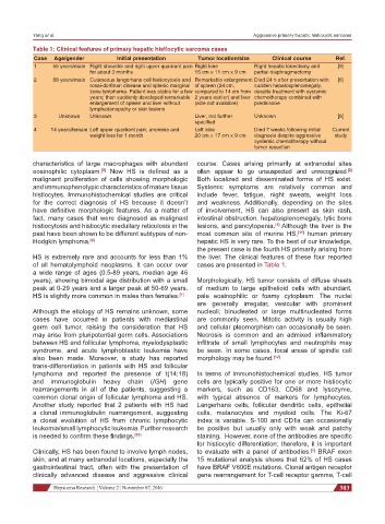
Table 1: Clinical features of primary hepatic histiocytic sarcoma cases
Case Age/gender Initial presentation Tumor location/size Clinical course Ref.
1 55 years/male Right shoulder and right upper quadrant pain Right lobe Right hepatic lobectomy and [9]
for about 3 months 15 cm × 11 cm × 9 cm partial diaphragmectomy
2 68 years/male Cutaneous langerhans cell histiocytosis and Remarkable enlargement Died 24 h after presentation with [6]
rosai-dorfman disease and splenic marginal of spleen (24 cm, sudden hepatosplenomegaly,
zone lymphoma. Patient was stable for a few compared to 14 cm from despite treatment with systemic
years; then suddenly developed remarkable 2 years earlier) and liver chemotherapy combined with
enlargement of spleen and liver without (size not available) prednisone
lymphadenopathy or skin lesions
3 Unknown Unknown Liver, not further Unknown [8]
specified
4 14 years/female Left upper quadrant pain, anorexia and Left lobe Died 7 weeks following initial Current
weight loss for 1 month 20 cm × 17 cm × 9 cm diagnosis despite aggressive study
systemic chemotherapy without
tumor resection
characteristics of large macrophages with abundant course. Cases arising primarily at extranodal sites
eosinophilic cytoplasm. Now HS is defined as a often appear to go unsuspected and unrecognized.
[2]
[8]
malignant proliferation of cells showing morphologic Both localized and disseminated forms of HS exist.
and immunophenotypic characteristics of mature tissue Systemic symptoms are relatively common and
histiocytes. Immunohistochemical studies are critical include fever, fatigue, night sweats, weight loss
for the correct diagnosis of HS because it doesn’t and weakness. Additionally, depending on the sites
have definitive morphologic features. As a matter of of involvement, HS can also present as skin rash,
fact, many cases that were diagnosed as malignant intestinal obstruction, hepatosplenomegaly, lytic bone
histiocytosis and histiocytic medullary reticulosis in the lesions, and pancytopenia. Although the liver is the
[1]
past have been shown to be different subtypes of non- most common site of murine HS, [11] human primary
Hodgkin lymphoma. [9] hepatic HS is very rare. To the best of our knowledge,
the present case is the fourth HS primarily arising from
HS is extremely rare and accounts for less than 1% the liver. The clinical features of these four reported
of all hematolymphoid neoplasms. It can occur over cases are presented in Table 1.
a wide range of ages (0.5-89 years, median age 46
years), showing bimodal age distribution with a small Morphologically, HS tumor consists of diffuse sheets
peak at 0-29 years and a larger peak at 50-69 years. of medium to large epithelioid cells with abundant,
HS is slightly more common in males than females. [1] pale eosinophilic or foamy cytoplasm. The nuclei
are generally irregular, vesicular with prominent
Although the etiology of HS remains unknown, some nucleoli; binucleated or large multinucleated forms
cases have occurred in patients with mediastinal are commonly seen. Mitotic activity is usually high
germ cell tumor, raising the consideration that HS and cellular pleomorphism can occasionally be seen.
may arise from pluripotential germ cells. Associations Necrosis is common and an admixed inflammatory
between HS and follicular lymphoma, myelodysplastic infiltrate of small lymphocytes and neutrophils may
syndrome, and acute lymphoblastic leukemia have be seen. In some cases, focal areas of spindle cell
also been made. Moreover, a study has reported morphology may be found. [12]
trans-differentiation in patients with HS and follicular
lymphoma and reported the presence of t(14;18) In terms of immunohistochemical studies, HS tumor
and immunoglobulin heavy chain (IGH) gene cells are typically positive for one or more histiocytic
rearrangements in all of the patients, suggesting a markers, such as CD163, CD68 and lysozyme,
common clonal origin of follicular lymphoma and HS. with typical absence of markers for lymphocytes,
Another study reported that 2 patients with HS had Langerhans cells, follicular dendritic cells, epithelial
a clonal immunoglobulin rearrangement, suggesting cells, melanocytes and myeloid cells. The Ki-67
a clonal evolution of HS from chronic lymphocytic index is variable. S-100 and CD1a can occasionally
leukemia/small lymphocytic leukemia. Further research be positive but usually only with weak and patchy
is needed to confirm these findings. [10] staining. However, none of the antibodies are specific
for histiocytic differentiation; therefore, it is important
Clinically, HS has been found to involve lymph nodes, to evaluate with a panel of antibodies. BRAF exon
[1]
skin, and at many extranodal locations, especially the 15 mutational analysis shows that 62% of HS cases
gastrointestinal tract, often with the presentation of have BRAF V600E mutations. Clonal antigen receptor
clinically advanced disease and aggressive clinical gene rearrangement for T-cell receptor gamma, T-cell
Hepatoma Research ¦ Volume 2 ¦ November 07, 2016 303