
Page 311 - Read Online
P. 311
Yang et al. Aggressive primary hepatic histiocytic sarcoma
A A
B B
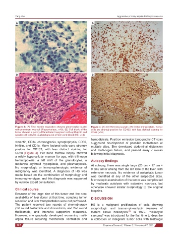
Figure 3: (A) Fine needle aspiration showed pleomorphic nuclei Figure 4: (A) CD163 immunostain; (B) CD68 immunostain. Tumor
with prominent nucleoli (Papanicolaou, ×40); (B) Cell block of the cells are strongly positive for CD163, with less distinct staining for
tumor showed a poorly differentiated neoplasm with epithelioid and CD68 (×10)
spindle cell features in a background of fibrin and blood (HE, ×40)
hemodialysis. Positron emission tomography CT scan
vimentin, CD34, chromogranin, synaptophysin, CD56, suggested development of possible metastases at
inhibin, and CD1a. Many lesional cells were strongly multiple sites. She developed abdominal distension
positive for CD163, with less distinct staining for and multi-organ failure, and passed away 7 weeks
CD68 [Figure 4]. Her bone marrow biopsy showed following initial diagnosis.
a mildly hypercellular marrow for age, with trilineage
hematopoiesis, a left shift of the granulocytes, a Autopsy findings
moderate erythroid hyperplasia, and plasmacytosis. At autopsy, there was single large (20 cm × 17 cm ×
No morphologic or immunophenotypic evidence of 9 cm) tumor arising from the left lobe of the liver, with
malignancy was identified. A diagnosis of HS was extensive necrosis. No evidence of metastatic tumor
made based on the combination of morphology and was identified at any of the other suspected sites.
immunophenotype, and this diagnosis was supported Microscopic examination of the tumor was complicated
by outside expert consultation. by moderate autolysis with extensive necrosis, but
otherwise showed similar morphology to the original
Clinical course biopsies.
Because of the large size of this tumor and the non-
availability of liver donor at that time, complete tumor DISCUSSION
resection and liver transplantation were not performed.
The patient received two rounds of chemotherapy HS is a malignant proliferation of cells showing
(1st round ifosfamide and doxorubicin, and 2nd round morphologic and immunophenotypic features of
thalidomide) and intensive supportive measures. mature tissue histiocytes. In 1970, “histiocytic
[7]
However, she gradually developed worsening multi- sarcoma” was introduced for the first time to describe
organ failure requiring mechanical ventilation and a collection of malignant tumor cells with histologic
302 Hepatoma Research ¦ Volume 2 ¦ November 07, 2016