
Page 310 - Read Online
P. 310
Yang et al. Aggressive primary hepatic histiocytic sarcoma
A A
B B
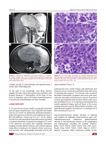
Figure 1: Abdominal magnetic resonance imaging (A: coronal; Figure 2: (A) Tumor cells comprising of primarily epithelioid cells
B: axial) revealed an 18.3 cm large heterogeneous enhancing with abundant eosinophilic cytoplasm, pleomorphic nuclei with open
mass with cystic components arising from the left hepatic lobe with vesicular chromatin and prominent nucleoli; (B) Mitotic figures were
associated mass effect easily found in the tumor. HE, ×40
multiple rounds of chemotherapy and passed away 7 upper quadrant [Figure 1].
weeks after initial diagnosis.
Laparoscopic liver needle biopsy was performed and
To the best of our knowledge, only three primary showed a tumor of primarily epithelioid cells with areas
hepatic HS cases have been previously reported in the of spindling also present. The lesional cells contained
English literature. [4-6] Recognition of this type of rare abundant eosinophilic cytoplasm and nuclei with open
tumor is important, due to its limited responsiveness to vesicular chromatin and prominent nucleoli. Prominent
conventional chemotherapy and high mortality. nuclear pleomorphism and elevated mitotic index were
also identified [Figure 2]. A subsequent perihepatic fine
CASE REPORT needle aspiration biopsy and cell block of the tumor
similarly showed a poorly differentiated neoplasm with
A 14-year-old previously healthy young girl presented epithelioid and spindle cell features in a background of
with left upper quadrant pain, loss of appetite and weight fibrin and blood [Figure 3].
loss for one month. An abdominal ultrasound showed a
large heterogeneous solid and cystic appearing mass in Immunohistochemical studies showed no staining
the mid abdomen, measuring 18.3 cm × 10 cm, arising with markers for epithelial, lymphomatous, germ cell,
from the liver with associated mass effect and surrounding rhabdomyosarcoma, melanoma, gastrointestinal
increased vascularity. Computed tomography (CT) scan stromal or neuroendocrine tumors, or for Langerhans
of the abdomen and pelvis and magnetic resonance cell histiocytosis, including CK cocktail, CAM5.2,
imaging revealed similar findings, with marked regional EMA, CD15, PAX-5, CD45, CD43, CD3, CD5, ALK1,
mass effect, including on the hepatic inferior vena cava Factor XIIIa, CD31, CD23, CD35, SALL-4, CD117,
and stomach, with extracapsular extension into the left desmin, myogenin, SMA, melanoma cocktail, S-100,
Hepatoma Research ¦ Volume 2 ¦ November 07, 2016 301