
Page 303 - Read Online
P. 303
Alves et al. Hepatocarcinoma with mediastinal metastasis
Non-invasive diagnosis is made by imaging A B
techniques, such as computed tomography and/
[4]
or magnetic resonance imaging, based on the
vascular findings for these tumors, which exhibit a
hypervascular pattern during the arterial phase and a
washout pattern during the portal venous or delayed
phase. Such radiological characteristic occurs in
a small number of 1-2 cm tumors. In these cases,
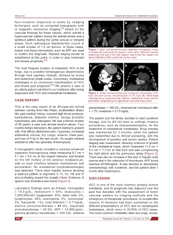
biopsy and tissue biomarkers, such as AFP, are used Figure 1: Axial contrast-enhanced computed tomography slices
to confirm the diagnosis. Disease staging should be at arterial (A) and portal (B) phases. Note tumor infiltration (arrow)
extending from part of the right hepatic lobe to the hilar region and
established at this point, in order to plan treatment tumor infiltration of the portal vein (arrow head)
and assess prognosis. [3,4]
A B
The most frequent location of metastatic HCC is the
lungs, due to possible hematogenous dissemination
through their capillary network, followed by bones
and abdominal lymph nodes. Conversely, mediastinal
metastasis is an uncommon manifestation of HCC
and shows poor prognosis. [5,6] We present a case of
an elderly patient admitted to our institution after being
diagnosed with HCC and mediastinal metastasis. Figure 2: Axial contrast-enhanced computed tomography (CT)
slice (A) and coronal reconstruction of CT scan (B). Note large
CASE REPORT heterogeneous hypervascular anterior mediastinal mass (arrow)
posteriorly compressing the right atrium and pulmonary artery
This is the case report of an 80-year-old retired phosphatase = 148 U/L; international normalized ratio
widower coming from São Paulo, southeastern Brazil. = 1.75; creatinine = 0.77 mg/dL.
His past medical history included high blood pressure,
dyslipidemia, diabetes mellitus, benign prostatic The patient and his family decided to start sorafenib
hypertrophy, and osteopenia. He was a former smoker therapy, and he did not want to undergo invasive
of 90 packs a year and denied alcohol abuse. Four procedures, such as chemoembolization, for the
months before admission, the patient started to present treatment of mediastinal metastasis. Drug therapy
with mild diffuse abdominal pain, hyporexia, increased was maintained for 3 months, when the patient
abdominal volume, dry cough, anterior chest pain, was readmitted due to clinical worsening, with the
and loss of 4 kg in the last mouth. He sought medical development of jaundice and severe ascites. Patient
assistance after two episodes of hemoptysis. staging was reassessed, showing evidence of growth
of the mediastinal mass, which measured 11.0 cm ×
A tomographic study revealed a contrast-enhanced 6.3 cm × 7.7 cm at that time and was compressing
expansive heterogeneous mass measuring 9.7 cm × the right atrium and the pulmonary artery [Figure 2].
5.1 cm × 5.6 cm at this largest diameter and located There was also an increase in the size of hepatic solid
on the left midline of the anterior mediastinum, lesions and in the extension of thrombosis. AFP levels
with no clear interface between mediastinum and reached 45,000 ng/mL. It was decided to discontinue
pericardium. No mediastinal lymphadenomegaly chemotherapy with sorafenib, and the patient died 1
was detected. There were hepatic lesions showing month after readmission.
a washout pattern in segments V, VI, VII, VIII and IV
and protruding toward the hepatic hilum. Evidence of DISCUSSION
splenic vein thrombus was found [Figure 1].
HCC is one of the most common primary tumors
Laboratory findings were as follows: hemoglobin worldwide, and its prognosis has improved over the
= 10.5 g/dL; hematocrit = 34%; leukocytes = past few decades with the assessment of tumor
7,500.000/mm (segmented: 64%, band cells: 0%, vascular pattern by imaging methods and the
3
lymphocytes: 28%, eosinophils: 5%, monocytes: emergence of therapeutic procedures. A considerable
3%, basophils: 1%); total bilirubin = 0.7 mg/dL; amount of literature has been published on the
alanine aminotransferase = 48 U/L; aspartate different presentations of HCC and on extrahepatic
aminotransferase = 40 U/L; AFP = 14,000 ng/mL; metastases, which occur in 30-50% of HCC cases.
gamma-glutamyl transferase = 350 U/L; alkaline The most common metastatic sites are lungs, bones,
294 Hepatoma Research ¦ Volume 2 ¦ October 21, 2016